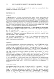
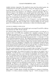
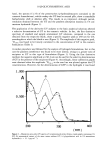
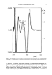
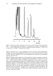
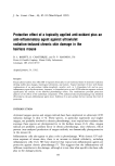
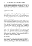
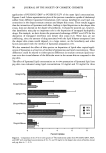
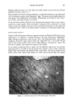
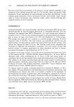
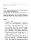
IMAGE ANALYSIS 103 Kligman rabbit ear assay (4). In the rabbit ear model, sample A received a score of 0 and sample B received a score of ! +. The test agents were blind coded and applied in a randomized manner on the upper back in five-centimeter squares using Dermapore © tape. The agents were liberally applied to each square, with reapplication on Mondays, Wednesdays, and Fridays for four consec- utive weeks, providing 28 days of occlusion. At the end of four weeks, FB slides were obtained and evaluated under a stereo micro- scope by an expert grader. These interpretive evaluations were conducted in a blind manner using the standard four-point scale mentioned previously. In this paper the "interpretive score" refers to the expert grader's evaluation of a follicular biopsy slide. DIGITAL IMAGE ANALYSIS Images of follicular biopsy slides are captured through an Olympus SZH light micro- scope with a ! x objective, overhead fiberoptic ring light illumination (Highlight © 3000, Olympus Corporation, Lake Success, NY), two polarizing filters, and a high- resolution Nuvicon © video camera (Series 68, Dage-MTI Inc.). An Intel © PC-AT system with a Joyce-Loebl © vision card set (Mini Magiscan, Compex Inc., Mars, PA) was used to digitize and analyze the captured video image. In our system a polarizing filter is placed over the fiberoptic light source and another polarizing filter is attached to the microscope lens. Figures 2 and 3 show a follicular biopsy slide under regular and polarized light, respectively. The horny impactions are barely visible under regular light (Figure 2), while under polarized light they appear Figure 2. A follicular biopsy slide viewed under regular illumination

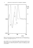
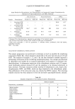
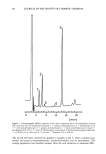
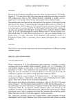
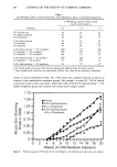
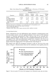
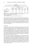
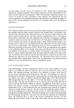
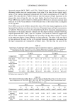
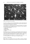
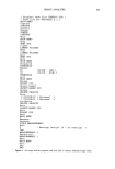
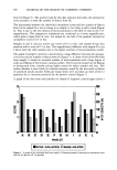
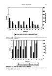
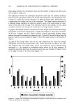
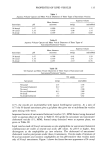
104 JOURNAL OF THE SOCIETY OF COSMETIC CHEMISTS L Figure 3. The same follicular biopsy as in Figure 2 viewed under polarized illumination. Note the contrast between the horny impactions and the background. more intense, standing out from the background (Figure 3). This contrast between the impactions and the background allows the samples to be evaluated by image analysis for total number and size distribution of impactions. A task (program) for evaluating FB slides under polarized illumination was developed that automatically counts the number, area, and size distribution of horny impactions. An IA task consists of the following basic steps: 1. Image capture (digitizing) 2. Segmentation 3. Object detection 4. Measurement/data storage 5. Analysis The task automatically controls what objects to evaluate (based on intensity range), what type of editing to perform, and what measurements to make and record. An example of a task used to evaluate follicular biopsy slides is illustrated in Figure 4. Image analysis data was obtained from the FB slides of the 12 panelists for each test sample. The follicular biopsies were evaluated at 15 x, giving a field of view 9.4 mm x 10.0 mm. The measurements are in units of pixels for comparative purposes. RESULTS AND DISCUSSION When looking at the individual data from each panelist for sample A (cleansing masque), we see a relationship between the interpretive scores and the number of objects
Purchased for the exclusive use of nofirst nolast (unknown)
From: SCC Media Library & Resource Center (library.scconline.org)