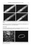
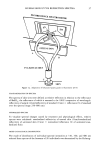
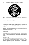
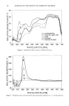
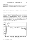
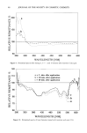
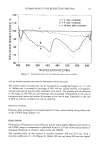
2 JOURNAL OF THE SOCIETY OF COSMETIC CHEMISTS In this study, we used two confocal microscopes to study the surface aspect and internal structure of human hair. The tandem-scanning reflected-light microscope (TSM), in- vented by Petran eta/. (3), gave high-resolution images of the surface, and was used to view and quantify the effects of cosmetic treatments, swelling and stretching on the topography of cuticular cells. Internal structures were visualized using a confocal laser scanning microscope (CLSM). Fine details of the cortex and medulla were identified by the use of a fluorochrome. MATERIALS AND METHODS HAIR SAMPLING Freshly plucked hair and eyelash were simply collected on the authors at the time of the experiment. In all other cases, commercially available Caucasian virgin brown hair was used in standardized conditions. HAIR TREATMENTS (4) A permanent wave treatment was comprised of two steps: reduction and oxidation. Reduction was done with ammonium thioglycolate (1 M), adjusted at pH 9 with ammonia, for 30 minutes at 30øC. Hair was rinsed and fixed with a 2.5% hydrogen peroxide solution adjusted at pH 3 with hydrochloric acid. Finally, samples were rinsed with tap water. Bleaching was performed by immersing virgin hair in hydrogen peroxide (6%)/sodium persulfate (10%) solution, adjusted to pH 10 with ammonia for 45 minutes at 35øC, and then thoroughly rinsing with tap water. Swelling was obtained by immersing virgin hair in either deionized water or 8 M urea at room temperature for ten minutes. STAINING WITH FLUOROCHROMES 10 mg of virgin hair were immersed for one hour in 5 ml of 0.05% solutions of rhodamin B in water or octadecyl-rhodamin in ethanol/water (9:1) at 60øC. The excess of fluorochrome was removed by dipping hairs for ten seconds in tetrachloroethylene the hair was then dried in an oven at 105øC overnight. BASIC PRINCIPLE OF THE CONFOCAL MICROSCOPE Whereas the classic light microscope homogeneously illuminates a large area within the sample, the confocal microscope acts with a focused beam of light. The reflected light at the focused point does go through a pinhole in front of a detector (TV camera, photo multiplier). Out-of-focus reflections cannot pass the pinhole and consequently are not imaged. An image is generated at the focal plane by scanning the light spot in X and Y directions with either rotating mirrors or a Nipkow disc. The image does not contain any information from above and beneath the focal plane and is called an optical section.

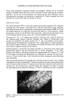
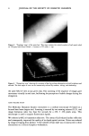
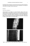
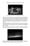
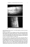
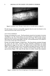
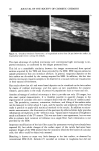
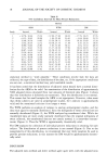
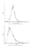
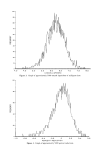
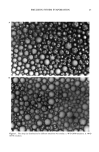
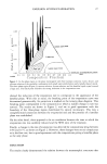
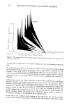
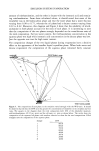
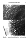
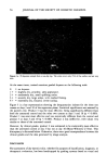
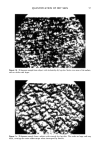
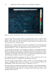
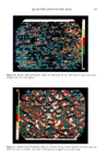
CONFOCAL 3D RECONSTRUCTION OF HAIR 3 Thick semi-transparent specimens (almost all biological tissues) can be vertically scanned. In-depth optical sections are then stacked for volume rendering by computer treatment. At the end of the procedure, the sample has not been destroyed. As no blurring spoils the image, the resolution is improved (0.25 !xm) compared with that obtained by conventional light microscopy (0.6 I•m). HAIR SURFACE STUDY The Tracor-Northern TSM is a real-time confocal microscope equipped with a Nipkow scanning disk. The sample is illuminated by a 100-watt mercury-arc lamp. Reflected light from the focal plane is captured by a monochrome video camera (COHU-0.02 lux) and rapidly digitized via 4-data flow processors (PC-Oeil) on a microcomputer. Image processing is carried out by PC-Oeil, which provides a 3D reconstruction based on up to 256 stacked optical sections using 3D + software (MISIS-Tracor Europa). This gives two 512 x 512 image arrays coded on 256 gray levels, which are known as the "reflection map" (Figure 1) and the "topologic map" (Figure 2). Respectively, they provide the brightness and spatial position (X, Y, and Z coordinates) of each pixel. Combining the two maps gives a "perspective view" image (Figure 3), which reproduces the cylindrical shape of the hair. Line profiles can be generated interactively and provide local surface roughness parameters (see below). Freshly-plucked or treated hair was simply attached to a microscope slide coated with double-sided adhesive tape and observed with no further preparation. A dry 100X objective was selected, providing a resolution of approximately 0.25 I•m in X, Y, and Z. 3D images containing full information were obtained by stacking 100 optical sec- tions in 0.1-1•m vertical steps. Noise reduction by frame averaging or gray processing was unnecessary. Hair was stretched between two parallel jaws moved by an electric motor, thus avoiding manipulation during the experiment. The hair was glued to the jaws using cyanoacrylate resin. Twenty optical sections in 0.5-1•m vertical steps were collected at each step of the extension for 3D reconstruction each 3D image thus obtained was made to overlap the preceding ones by interactive shifting in X and Y coordinates in order to examine Figure I. "Reflectance map" of a natural brown hair. Local changes in brightness can be evaluated on a 256 gray scale range.
Purchased for the exclusive use of nofirst nolast (unknown)
From: SCC Media Library & Resource Center (library.scconline.org)