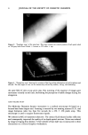
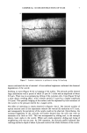
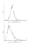
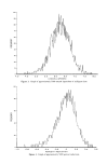
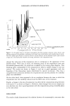
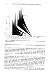
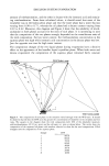
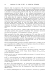
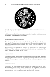
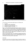
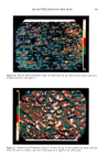
4 JOURNAL OF THE SOCIETY OF COSMETIC CHEMISTS : Figure 2. "Topologic map" of the same hair. This map contains the vertical position of each pixel coded on 256 gray levels from O-black = bottom to 255 white = top. Figure 3. "Perspective view" showing the curvature of hair by pooling information on both brightness and altitude. The best angle of view can be interactively selected by rotation, tilting, and shadowing. the same field of view at any given step. Fast scanning of the sequence of images gave movement virtually in real time, facilitating the perception of subtle changes during the elongation. HAIR VOLUME STUDY The Molecular Dynamics Sarastro instrument is a confocal microscope (5) based on a focused laser beam (argon-ion). Scanning is ensured by two rotating mirrors (X,Y), and image formation takes less than five seconds for a 256 x 256 pixels array. This microscope is used to acquire fluorescence signals. We selected a 60X oil-immersion objective. The contact fluid reduced surface reflection and consequently improved the quality of in-depth optical sections. Noise was reduced by image averaging (four frames). A full cylinder of hair shaft was reconstructed in three dimensions on a Silicon Graphics workstation.

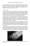
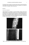
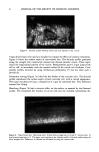
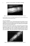
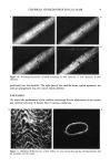
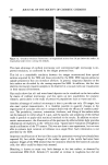
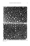
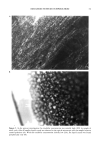
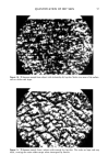
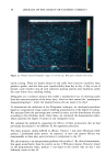
CONFOCAL 3D RECONSTRUCTION OF HAIR 5 The fluorescent hairs were glued to microscope slides coated with double-sided adhesive tape. Between 40 and 100 optical sections were stored, depending on the diameter of the hair shaft, for complete volume reconstruction. RESULTS HAIR SURFACE EXPERIMENTS Freshly plucked hair was imaged in its natural environment. Figure 4a shows sweat droplets attached to the free border of cuticular cells. The joints between the successive optical sections were invisible and the border lines of the cuticle were finely drawn with no interruption. The confocal image was comparable to an SEM picture (Figure 4b). Figure 5 shows natural deposits of sebum and dust particles on an eyelash. Sebum filled the spaces between adjacent cuticular cells, facilitating dust deposition. Neither sweat nor sebum could be observed in conventional SEM. Figure 4. Freshly plucked hair: a) confocal image showing sweat droplets b) "cleaned" SEM image.
Purchased for the exclusive use of nofirst nolast (unknown)
From: SCC Media Library & Resource Center (library.scconline.org)