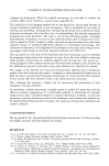
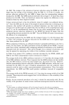
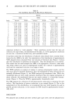
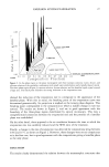
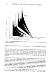
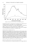
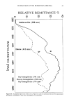
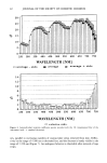
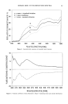
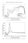
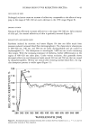
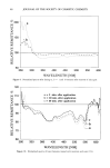
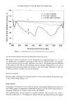
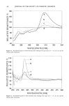
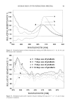
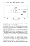
j. Soc. Cosmet. Chem., 44, 35-52 (January/February 1993) Fluorescence-free UV/VIS reflection spectra of human skin G. SAUERMANN, A. HERPENS, D. DREWES, A. GRIMMERT, B. AUGUST, and U. HOPPE, Paul Gerson Unna Research Center of Beiersdorf AG, D-2000 Hamburg, and Fachhochschule fiir Chemieingenieurwesen, D-2000 Hamburg, Germany. Synopsis A fluorimeter operated in synchronous mode was used to record fluorescence-free UV/VIS reflection spectra of human skin. BaSO 4 or Teflon served as reflection standards. The variation coefficient of the spectral data showed a maximum around 350 nm. The relative remittance (normalized spectra) increased dramatically after cyanoacrylate stripping around 305 nm equivalent with loss of UV-absorption. Increase of moisture level by occlusion or washing of skin caused a similar but reversible change, washing with surfactants was provocing changes lasting for more than 24 hours. Exaggerated use of soap and surfactant bars drastically enlarged remittance around 300 nm. INTRODUCTION UV/VIS radiation impinging on the skin surface will be only partially directly reflected. About 5% of the total radiation energy is reflected from the surface (1). The remaining radiation entering the stratum corneum will be reflected, diffracted, and refracted, processes that are described in total as scattering (2). Therefore, the light beam will lose its original direction and tend to gain a more diffuse distribution within the skin. During its chaotic way through tissue, light will be diminished by endogenous and exogenous chromophores absorbing at different wavelengths: the longer the optical pathway, the more the loss by absorption. With increasing wavelengths, irradiation reaches deeper layers of the skin (3). A fraction of the incident radiation of different wavelengths, scattered from different penetration depths, will reach the skin surface again (= diffuse reflection). Depending upon the quality and quantity of the chromopheres that rays encounter on their penetration path, the recorded spectra will show more or less intense absorption peaks. The whole level and general shape of the spectra will be determined by the scattering properties of the skin getting increasingly turbid or less permeable with shorter wavelengths (1). To standardize spectra, the particular influences of lamps, optical geometry, and skin probes have to be eliminated. This is optimally achieved by producing relative spectra that show the diffuse remittance of skin in percentages of the reflectivity of a reflection standard like BaSO 4 or Teflon (reflection close to 100%, irrespective of wavelength). 35

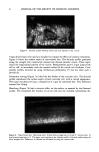
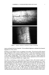
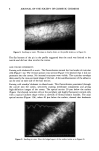
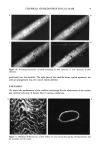
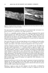
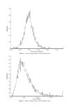
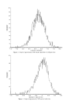
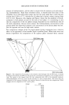
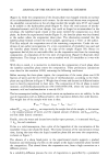
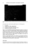
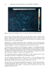
36 JOURNAL OF THE SOCIETY OF COSMETIC CHEMISTS Traditionally the reflection of monochromatic light is recorded using an integrating sphere connected to a photometer (4-6). Such an instrumental array does not allow discrimination of reflection from fluorescence: when using a UV-photometer the re- flected light after interaction with skin is no longer limited to a controlled wavelength. Fluorescence contributes considerably to the recorded signal when skin is irradiated with UV (1) because the epidermis contains a series of UV-absorbing fluorescent molecules: aromatic aminoacids, NADH, flavines, porphyrines, possibly Maillard products, and lipofuscin (7). To record only reflection and eliminate fluorescence of different wave- lengths, a commercial fluorimeter with monochromators in the excitation as well as in the emission pathway has been used and operated in the so-called synchronous mode, i.e., both monochromators are moved synchronously through the whole spectrum with a wavelength difference of zero. To measure optical data at arbitrarily chosen skin sites, light guides transporting light from the instrument to the skin, and after interaction with the skin back to the instrument for further analysis, are preferentially chosen. The moisture content of the excised horny layer is steadily related to the relative humidity of the environment (8). A frequently used technique uses electrical capacitance to measure relative water content of the skin (9). We found that application of dry silica gel under occlusive dressing decreases electrical capacitance measured with an appro- priate instrument (Corneometer, Courage-Khasake, Cologne, Germany) and therefore probably skin moisture content (t0). This technique to reduce skin moisture content was used to get optical data on dry skin. Contact of skin with water elutes amino acids, urocanic acid, lactic acid, etc., from the horny layer (11). UV-spectrometric measurements of such aqueous extracts show ab- sorption maxima around 260-280nm. Quantification and identification of the main components as urocanic acid and tyrosine can be achieved by HPLC (t0). EXPERIMENTAL DETAILS INSTRUMENTS To measure reflection spectra a fluorimeter (LS 50, Perkin-Elmer) was operated in synchronous mode with a wavelength (h.) difference between excitation and emission monochromator (Ah. = 0 nm). To avoid overloading of the photomultiplier, filters were introduced into the excitation pathway: attenuator t %. Both arms of a bifurcated quartz guide (UV-type, Schott, Mainz, Germany) were installed into the fiber-optic holder (Figure ta). The end, C, of the fiber-optic should contain the individual fibers coming from branches A and B in a statistical or optimally evenly distributed manner to avoid as far as possible specular reflection (Figure lb). C is surrounded by a transparent and stiff disc of plastic (radius: 3 cm). Transparency is necessary to precisely select skin areas a disc should minimize changes of dermal blood volume caused by pressure. REFLECTION STANDARDS Commercially available standards of BaSO 4 and Teflon were used.
Purchased for the exclusive use of nofirst nolast (unknown)
From: SCC Media Library & Resource Center (library.scconline.org)