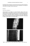
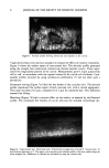
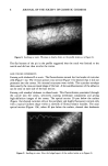
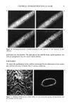
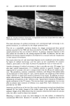
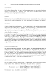
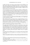
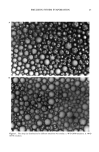
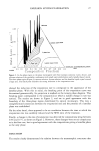
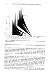
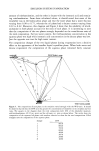
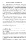
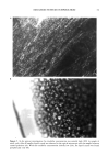
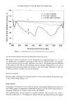
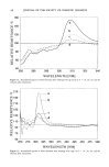
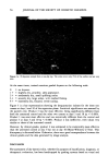
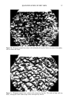
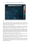
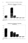
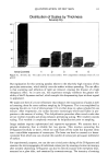
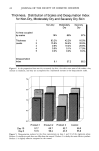
8 JOURNAL OF THE SOCIETY OF COSMETIC CHEMISTS Figure 8. Swelling in water. Waviness is clearly drawn on the profile (scales as in Figure 6). The flat bottom of the pit in the profile suggested that the crack was limited to the cuticle and did not thus involve the cortex. HAIR VOLUME EXPERIMENTS Staining with rhodamin B in water. The fluorochrome stained the free border of cuticular cells (Figure 1 la). The vertical optical cross section (Figure 1 lb) showed that it did not penetrate into the cortex. No internal structures were visible. The cuticular envelope drew exactly the cross-sectional shape of the hair. A fair autofluorescence of the adhesive can be seen on each side of the hair section. Staining with octadecyl rhodamin in ethanol:water. This fluorochrome penetrated through the cuticle into the cortex, selectively staining membrane components and giving high-definition images of the cortex. The optical section 20 Ixm below the surface (Figure 12a) showed cuticular cells at the periphery and highly fluorescent cortical cells with a typical fusiform shape within a network of fibrous keratin bundles. The axial optical section (Figure 12b), taken 40 Ixm below the surface, showed that rhodamin Figure 9. Swelling in urea. Note the bulged aspect of the surface (scales as in Figure 6).

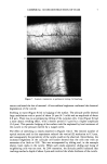
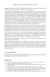
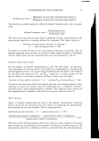
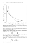
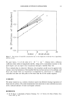
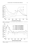
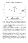
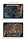
CONFOCAL 3D RECONSTRUCTION OF HAIR 9 Figure 10. Stretching experiment: a) before stretching b) 10% extension c) 15% extension d) 20% extension. penetrated into the medulla. The right part of the medulla shows typical septation the cortical arrangement was even more clearly defined. DISCUSSION We tested the performance of the confocal microscope for the observation of the surface and internal structure of human hair in various conditions. Figure 11. Rhodamin B fluorescence: a) hair surface b) cross section showing that the fluorochrome did not penetrate into the cortex.
Purchased for the exclusive use of nofirst nolast (unknown)
From: SCC Media Library & Resource Center (library.scconline.org)