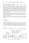
282 JOURNAL OF THE SOCIETY OF COSMETIC CHEMISTS felting of wool (14). In the case of the aliphatic alcohol of longer chain, the degradative reaction is confined to the surface of the wool. Taki et al. (3) and Taki (4) reported that descaled wool fibers were obtained without any damage to cortical cells when potassium hydroxide solution in dehydrated 1-butanol was used. This method, however, has not been applied to human hair, albeit it is one of the most significant methods for removing cuticle cells from human hair. The objective of the present study is to investigate the process of removing several layers of cuticle in human hair by applying Taki's methods using KOH/1-butanol (3). Detailed morphological changes in cuticle under the various conditions of chemical treatment are discussed by means of scanning electron microscopy (SEM) and transmission electron microscopy (TEM). We fhrther observe the change in the amino acid residues by the treatment based on Fourier transform infrared spectros- copy (FT-IR). MATERIALS AND METHODS SPECIMENS Human hair from a ten-year-old Japanese girl was used. The bundles of hair fibers were cut to a length of 80 mm. CHEMICAL ISOLATION OF CUTICLE CELLS About 3 g of the hair was washed in 100 cm 3 aqueous solution of 10% (w/v) sodium lauryl sulfate and then rinsed thoroughly in distilled water. Specimens were then washed in excess volume of acetone and dried. KOH/1-butanol solution was dehydrated by anhydrous K2CO 3 prior to use. Chemical isolation of the cuticle was carried out in the solution of 5.0 or 6.0% KOH/1-butanol (hair:liquor ratio was l:250 w/v) at 50øC for various treatment times. In Table I, five kinds of specimens with different degrees of chemical treatment are listed. After the chemical treatment, the specimens were ira- Table I Experimental Conditions for Removing Cuticle Cells From Human Hair KOH Descaled concentration hair (%) Treatment time and temperature Shaking condition DS-1 5 DS-2 5 DS-3 5 DS-4 5 DS-2' 6 50øC, 6 hr--• room temperature, 16 hr 50øC, 5 hr-• room temperature, 12 hr--- 50øC, 5 hr (50øC, 9 hr--- room temperature, 16 hr) 3 times repeat* (50øC, 9 hr--• room temperature, 16 hr) 3 times repeat --- 50øC, 9 hr* 50øC, 5 hr-• room temperature, 12 hr-• 50øC, 5 hr Manual stirring once every hour Rapid stirring (170 stroke/min) constantly Rapid stirring (170 stroke/min) constantly Rapid stirring (170 stroke/min) constantly Rapid stirring (170 stroke/min) constantly * The solution was renewed each day.

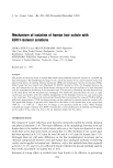
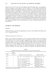
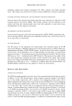
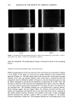
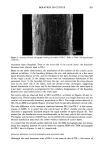
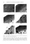
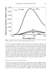
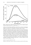
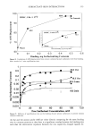
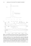
ISOLATION OF CUTICLE 283 mediately washed with ethanol containing 25% HC1, ethanol, and finally distilled water. The solution was renewed five times to remove KOH and 1-butanol completely. SCANNING ELECTRON MICROSCOPY AND TRANSMISSION ELECTRON MICROSCOPY Vertical views of the untreated and treated specimens were observed in a Hitachi S-4100 scanning electron microscope (SEM). The average diameter and the thickness of the cuticle cells from the cross-sectional views were also measured on more than thirty monofilaments of the respective specimens. TRANSMISSION ELECTRON MICROSCOPY Cross-sectional views of cuticle cells were observed by a JEOL 100SX transmission elec- tron microscope (TEM). Specimens were embedded in epoxy resin, and sections were cut on an ultra thin-cut microtome. FT-IR SPECTROSCOPY The IR spectra of the untreated and treated hairs were measured using an FT-IR microscope (Shimazu, IMS-800, Japan) and an FT-IR spectrometer in diffuse reflectance measurements (Shimazu, FTIR-8100M, Japan). To get the thin, flat section of exposed cortical cells, the monofilament was cut longitudinally on both sides. A transmission spectrum of 15 •m X 15 •m/square area of cortex was obtained with the FT-IR microscope (200 scans at 4 cm- • resolution) and an MCT detector. The spectra of the surface of the hair were obtained by scratching the surface of hair carefully with a blade. The scraped section was collected and diffuse reflectance measurements were carried out. The spectra analyzed were 200 scans at 4 cm- • resolution. In diffuse reflectance measurements, the Kubelka-Munk transformation was applied to obtain the final spectra. RESULTS AND DISCUSSION CHANGE IN THE SURFACE The SEM microphotographs of vertical views of the untreated and treated hair are shown in Figure 1 (a-d). The untreated hair clearly displays regularly arranged cells (Figure la). In the mildly treated hair, DS-l, however, the overlapping of the cuticle cells is rather ambiguous and some of the cuticle cells are torn off (Figure lb). The overlapping of the cells decreases significantly in DS-2 hair, resulting in a smoother surface (Figure lc). In the DS-3 hair, cuticle cells are not detected and the surface becomes gritty in texture. A similar observation is made with DS-4 hair (not shown in Figure 1). In Figure 2, the SEM micrograph of DS-2' hair treated in 6% KOH/1-butanol is shown. As the treatment condition of DS-2' is the same as that of DS-2 except that a higher KOH concentration was used, we duly compare an SEM microphotograph of DS-2' with that of DS-2 in Figure lc. The cuticle is still found, but the surface is not so smooth as that in DS-2. From the SEM microphotographs, it is difficult to determine how many cuticle
Purchased for the exclusive use of nofirst nolast (unknown)
From: SCC Media Library & Resource Center (library.scconline.org)