346 JOURNAL OF THE SOCIETY OF COSMETIC CHEMISTS

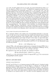
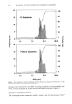
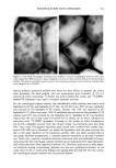
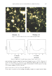
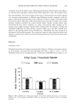
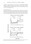
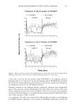
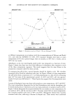
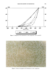
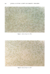
ENCAPSULATION INTO LIPOSOMES 347 3). Thus there was no binding of inulin to the surface of the liposomes that could yield an overestimation of EE. Furthermore, using this procedure, we were able to adequately separate free from encapsulated inulin (Figure 3). For these reasons, this method was selected for subsequent work. A disadvantage, however, is that liposome samples become contaminated with Ficoll © and are lost for use in further experiments. EVALUATION OF ENCAPSULATION EFFICIENCIES (EE) EEs obtained with various preparation methods and lipid concentrations are listed in Table I. RPE protocol 2 gave the highest EEs, with values ranging from 22-47%. These values are higher than EEs for inulin reported by other investigators (Table I) and compare well with those reported for other hydrophilic actives (Table II). Tables I and II also show wide variations in EE from one compound to another within a single preparation method. For example, liposomal gelonin prepared by RPE protocol 3 yielded EEs as high as 79% (Table II), whereas we found an EE of only 8% for liposomal inulin prepared by this method (Table I). Similar findings were noted earlier by Pidgeon et al. (24), who recommended that one always assay EE directly rather than relying on sucrose or carboxyfiuorescein data. Our results support this position. CONCLUSION The Ficoll © discontinuous density gradient method was found to be an excellent tool for separating free inulin from large, multilamellar liposome preparations. Using this method to determine encapsulation efficiencies (EE), we found that EE values approach- ing 40% were obtained by the reverse-phase evaporation (RPE) method, regardless of cholesterol level. The microstructure was shown to consist of large heterogeneous mul- tilamellar vesicles with diameters of 2-20 pro. At equal lipid concentration, much lower EE values (ca. 4%) were obtained for liposomes prepared by the film method. For inulin, comparison with other studies shows that EEs should be measured rather than inferred from systems having different compositions or preparation methods. ACKNOWLEDGMENTS We wish to thank Janet Burns for the cryo-TEM analysis, Brian Barford for assistance with the particle size measurements, and Dave Siegel for helpful discussions on liposome morphology. REFERENCES (1) K. Egbaria and N. Weiner, "Liposomes as a Topical Drug Delivery System Evaluated by In Vitro Diffusion Studies," in Cosmetic and Pharmaceutical Applications of Polymers, C. G. Gebelein, Ed. (Plenum Press, New York, 1991), pp. 215-224. (2) D. Yarosh, C. Bucana, C. Cox, L. Alas, J. Kibitel, and M. Kripke, Localization of liposomes containing a DNA repair enzyme in murine skin, J. Invest. Dermatol., 103, 461-468 (1994). (3) F. Bonina, L. Montenegro, C. La Rosa, F. Gasparri, and R. Leonardi, Comparison of different separative techniques in the quantitative determination of active compound enclosed in liposomal systems, Int. J. Cosmet. Sci., 16, 183-197 (1994).
Purchased for the exclusive use of nofirst nolast (unknown)
From: SCC Media Library & Resource Center (library.scconline.org)