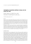
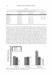
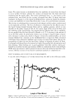
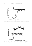
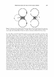
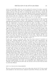
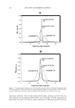
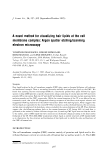
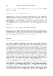
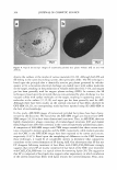
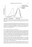
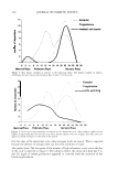
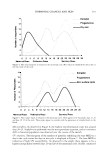
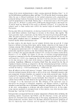
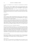
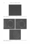
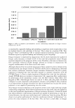
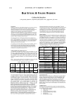
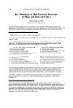
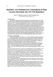
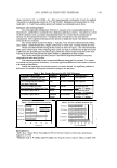
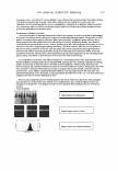
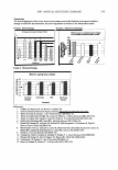
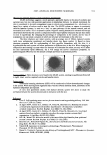
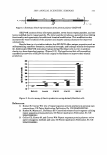
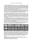
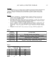
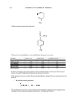
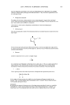
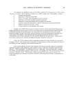
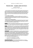
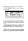
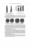
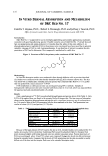
376 JOURNAL OF COSMETIC SCIENCE IN VITRO DERMAL ABSORPTION AND METABOLISM OF D&C RED No. 17 Camille T. Haynes, Ph.D., Robert L. Bronaugh, Ph.D. and Jeffrey J. Yourick, Ph.D. Office of Cosmetics and Colors, Food & Drug Administration, Laurel, MD 20708 Introduction D&C Red No. 17 is approved for use in externally applied drug and cosmetic applications, in amounts consistent with good manufacturing practice. Possible cosmetic uses of D&C Red No. 17 include skin and hair care preparations and suntan products ( 1 ). Concerns about the safety of this color additive ( 1-[ 4- phenylazophenylazo ]-2-naphthol (PAN) is the primary color constituent) have been raised due to potential metabolic cleavage of PAN to yield 4-aminoazobenzene. It was therefore of interest to examine the skin penetration of PAN and to determine if this compound is metabolized in viable skin. Figure 1: Structure of PAN, the primary color constituent of D&C Red No. 17 Methodology In vitro skin absorption studies were conducted in flow-through diffusion cells as previous described (2). Studies were conducted with either freshly obtained viable pig skin or human cadaver skin. The skin was dosed with a commercially available sunscreen product that contained D&C Red No. 17 ( 15 µg/rnl) that was spiked with a"tracer amount of 14 C-PAN. PAN metabolism in viable porcine skin was investigated using HPLC methods. Samples of homogenized viable porcine skin were extracted with ethyl acetate (1:2 vol:vol), which was separated from the aqueous layer and concentrated under a N2 stream. Results Only a small amount of 14 C-PAN was absorbed through human and porcine skin in 24 h (Table 1). Only 0.05% of the applied dose was found in the receptor fluid with human skin. The time course of absorption shows similar absorption between human and pig skin with a significant increase in pig skin absorption in the 24 h sample. Most of the PAN penetrating into the skin remained in the skin. Total penetration was similar in human (10.5 %) and porcine (13.2 %) skin. Table 1: PAN penetration in human and porcine skin after 24 hours (% total applied dose) Human Porcine Receptor fluid 0.05±0.0l 0.53±0.15 Skin content 10.5±1.7 12.6±1.2 Total applied 10.5±1.7 13.2±2.1 dose penetrated Wash 91.8±11.7 91.6±4.7 Recovery 102.4±11.1 104.2±4.9 Values are mean± SEM for human (n=7) and porcine (n=16) skin Because of the large amount of PAN found in the skin at 24 h, extended studies were conducted for 72 h (with a skin wash at 24 h) to determine if additional PAN would be absorbed (Table 2). Receptor fluid levels increased slightly with human skin and were now similar to pig skin values. Porcine skin levels of PAN were unchanged when 24 and 72 h values were compared. Human skin levels also did not decrease during the extended study. It appears that the levels of PAN penetrating into the skin are unable to readily diffuse out of the skin into the receptor fluid even in the extended absorption study.

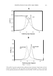
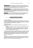
2005 ANNUAL SCIENTIFIC SEMINAR 377 Table 2: PAN penetration in human and porcine skin after 72 hours (% total applied dose) Human Porcine Receptor fluid 0.30±0.04 0.45±0.15 Skin content 17.1±4.4 12.2±2.2 Total applied 17.4±4.4 12.7±2.2 dose penetrated Wash 91.5±6.7 96.9±7.8 Recovery 108.9±7.6 109.5±9.3 Values are mean ± SEM for human (n=8) and porcine (n=4) skin Metabolism of PAN could only be examined in extracts from pig skin samples since the levels of radioactivity that were absorbed into the receptor fluid were extremely small. In spite of the fact that large amounts of radioactivity were present in the skin at 24 h, no metabolites of PAN were detected by HPLC analysis. Metabolism of PAN to 4-aminoazobenzene in skin would require the cleavage of an azo bond. We have previously shown that several other azo colors were significantly metabolized with azo bond cleavage in mouse, hairless guinea pig and human skin (3). However, these colors had better water solubility and were more readily absorbed than PAN. Human skin samples in the current study were not analyzed for metabolism since they were fipm non-viable cadaver skin. Conclusion The skin absorption of 14 C-PAN into the receptor fluid beneath the skin was significantly less than 1 % with either human or pig skin in both the 24 and 72 h studies. Substantial amounts of 14 C-PAN {10-17 %) penetrated into human and pig skin in 24 hr. Pig receptor fluid levels of PAN did not increase in the extended absorption studies suggesting that material in the skin may not be available for systemic absorption. Human receptor fluid levels of PAN did increase significantly at 72 h but the 72 h values were still low and similar to those with pig skin. No metabolism of PAN to 4-aminoazobenzene was observed in viable pig skin. References 1. Gottschalck, T. E. and McEwen, G. N., International Cosmetic Ingredient Dictionary and Handbook, 9'h Ed., The Cosmetic Toiletry and Fragrance Association, Washington DC, 2004. 2. Bronaugh, R.L. and S.W. Collier, Protocol for in vitro percutaneous absorption studies. In In Vitro Percutaneous Absorption: Principles, Fundamentals, and Applications, ed. By R.L. Bronaugh and H.I. Maibach, pp. 23 7-241. CRC Press, Boca Raton, 1991. 3. Collier, S. W, Storm, J.E., and Bronaugh, R. L., Reduction of azo dyes during in vitro percutaneous absorption, Toxicol. Appl. Pharmacol. 118, 73-79, 1993.
Purchased for the exclusive use of nofirst nolast (unknown)
From: SCC Media Library & Resource Center (library.scconline.org)