PHOTOCYTOTOXICITY OF TITANIUM DIOXIDE 537 White or lightly colored shades of inks were selected. The purchases were made between January 2006 and May 2010. All inks were explicitly labeled by the vendors for use as permanent makeup. GRAVIMETRIC DETERMINATION OF PERMANENT MAKEUP INKS’ PIGMENT CONTENT A 50-μl aliquot of permanent makeup ink was deposited onto a preweighed inorganic membrane fi lter (0.02-μm pore size, 10-mm diameter Whatman Inc., Clifton, NJ). The fi lter with deposited ink was weighed, and subsequently washed fi ve times under gentle vacuum using 50 μl of distilled water. The fi lter was then dried for three days in a ventilated oven operating at 35° ± 2°C. The fi lter was then weighed to obtain the weight of the dried pigment. The weight of the dried pigment and the initial weight of the permanent makeup ink were used to calculate the percentage of pigment in the permanent makeup ink. ISOLATION OF PIGMENTS FROM PERMANENT MAKEUP INKS One ml of permanent makeup ink was diluted with 3 ml of deionized water and centri- fuged at 85,000g (15°C) for one hour. (Optima L-90K ultracentrifuge, Beckman Coulter, Inc., Brea, CA). Sedimented pigments were washed twice by resuspension in 4 ml of deionized water and centrifugation as above. Pigments were then dried overnight under vacuum at 30°C. ELEMENTAL ANALYSIS OF PIGMENTS FROM PERMANENT MAKEUP INKS BY X-RAY FLUORESCENCE Each sample of pigment (approximately 200 mg) was mixed with paraffi n wax and pressed in a pellet die at 30 tons for fi ve minutes to form a standard pellet. X-ray fl uorescence mea- surements were made using a Bruker S4 wavelength dispersive X-ray fl uorescence spectrom- eter (Bruker AXS Inc., Madison, WI). The spectrometer sequentially searches for elements with atomic numbers from Na to U and adjusts the test conditions for each element to op- timize the detection sensitivity. A semiquantitative analysis was performed using the fl uo- rescence yield for each element and accounting for enhancements attributed to secondary excitation and absorption due to heavy elements. The semiquantitative analysis has a typical accuracy of 5%. The elements Al, Si, and Ti are reported as their most common oxides. DETERMINATION OF THE CRYSTALLINE PHASE OF TiO2 BY X-RAY DIFFRACTION Pigments isolated from permanent makeup inks were loaded onto a zero background holder and placed into a Phillips PW3020 diffractometer (Phillips Electronic Instru- ments, Inc., Mahwah, NJ) using Cu-Kα radiation at 40 KV and 30 mA. Scans were run over the range of 20° to 80° with a step size of 0.02° and a counting time of four hours. The crystalline phase of TiO2 was identifi ed using the powder diffraction fi le published by the International Centre for Diffraction Data (Newtown Square, PA). Once all phases were identifi ed, the amount of TiO2 in each was quantifi ed using a Rietveld refi nement for comparing the computed diffraction pattern with the observed diffraction pattern. CELL CULTURE Human skin fi broblasts (ATCC CRL-1634) were obtained from the American Type Culture Collection (Manassas, VA). Cells were cultured in Dulbecco’s modifi ed Eagle’s

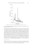
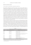
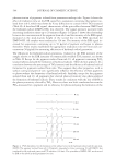
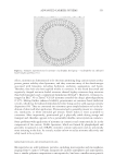
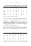
JOURNAL OF COSMETIC SCIENCE 538 medium, without phenol red, containing 10% fetal bovine serum, 50 μg/ml gentamicin, 4.5 mg/ml glucose, and 4 mM L-glutamine. All reagents used for cell culture were obtained from Invitrogen Corp., Carlsbad, CA. Cultures were incubated at 37°C in a humidifi ed atmosphere containing 5% CO2. IN VITRO ASSAY FOR CYTOTOXICITY AND PHOTOCYTOTOXICITY Fibroblasts were incubated for 18 hours with media containing either a permanent makeup ink or the pigment isolated from an ink. For treatment with a permanent makeup ink, a stock solution of the ink was prepared by dispersing the ink in deionized water us- ing a ten-second ultrasonic burst (Vibra-Cell VC250B sonicator, Sonics & Materials Inc., Danbury. CT). The stock solution was then heated for ten minutes at 100°C to minimize microbial contamination. Working solutions for treating fi broblasts were prepared by appropriately diluting stock solutions with media. Using the pigment content of each ink determined by gravimetric analysis, the level of treatment was expressed as the amount of pigment contained in the ink for each treatment and is given as μg of pigment per surface area of the fi broblast monolayer. For treatment with pigments, the pigments were dispersed in deionized water as described above and diluted in media to obtain working solutions used to treat fi broblasts. Following the 18-hour incubation in media containing a permanent makeup ink or pig- ment, fi broblasts were washed once with phosphate-buffered saline (PBS). Fibroblasts were then irradiated through freshly added PBS with 10 J/cm2 UVA radiation (320 nm– 400nm) combined with 45 J/cm2 visible light (400 nm–800 nm). Similar levels of UVA radiation and visible light would be received after exposure to the summer sun for 30 minutes (23). The source of UVA radiation and visible light was a 250-watt HITLite metal halide bulb (BLV Licht-und Vakuumteenik GmbH, Steinhöring, Germany) fi l- tered through glass. The emission spectrum of the light source was measured using an OL 754 UV-visible spectroradiometer (Optronic Laboratories Inc., Orlando, FL) and is shown in Figure 1. The spectral irradiance of the light source was found to be typically 6.3 × 10⁻³ W/cm2 UVA radiation and 2.8 × 10⁻² W/cm2 visible light. The emission of UVB radiation (280 nm–320 nm) from the light source was negligible (i.e., 1.1 × 10⁻⁷W/cm2). All irradiations were performed at 25° ± 3°C, and lasted approximately 25 minutes for simultaneous delivery of 10 J/cm2 UVA radiation and 45 J/cm2 visible light. To compen- sate for any inhomogeneity in the fi eld of illumination, uncovered samples were placed on a platform that rotated at 0.5 revolutions/min during irradiation. Sham-irradiated (i.e., dark control) samples were maintained at 25° ± 3°C in the dark. After irradiation, cells were removed from the dishes by trypsinization and plated into 60-mm Petri dishes (∼800 cells/dish). Four replicate dishes were plated for each treatment. The dishes were then incubated for 10–14 days to allow formation of cell colonies. Colonies were fi xed with methanol, stained with Giemsa stain, and counted. The average number of colonies observed after a treatment and the average number of colonies observed for cells receiving no treat- ment were used to calculate the percentage of cells surviving a treatment. Cytotoxicity was assessed using survival data for fi broblasts receiving treatment with an ink or a pigment alone (i.e., sham-irradiated samples), while photocytotoxicity was assessed from survival data for cells additionally exposed to light. A four-parameter logistic function (SigmaPlot 8, SPSS Inc., Chicago, IL) was used to fi t the data (i.e., % survival versus μg/cm2) and to determine the PD50 (dose of pigment which, in the presence of light, reduced survival by 50%).
Purchased for the exclusive use of nofirst nolast (unknown)
From: SCC Media Library & Resource Center (library.scconline.org)