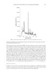
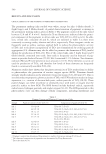
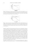
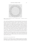
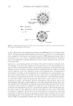
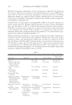
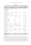
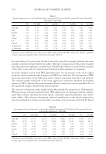
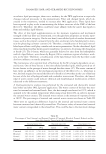
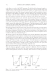
PHOTOCYTOTOXICITY OF TITANIUM DIOXIDE 541 nucleic acids, leading to photocytotoxicity (32–36). The crystalline form of TiO2 affects its effi ciency for generating ROS. Studies have demonstrated that TiO2 (anatase) is dramati- cally more photocatalytically active and photocytotoxic than the TiO2 (rutile) (24,33,37–39). We used X-ray diffraction to determine the crystalline phase of TiO2 in samples of TiO2 sold as anatase or rutile and in pigments isolated from permanent makeup inks (Table II). The sample of TiO2 sold as anatase was predominately anatase but contained 1.8 % TiO2 (rutile). One of the samples sold as TiO2 (rutile) contained 4.7% TiO2 (anatase), while no anatase was detectable in the other sample of TiO2 (rutile). Anatase was the primary crys- talline form of TiO2 found in the pigments isolated from six of the permanent makeup inks (inks 1, 2, 3, 6, 7, and 10). Ink 5 contained comparable amounts of TiO2 (anatase) and TiO2 (rutile). Ink 4, though predominately TiO2 (rutile), contained 0.6% TiO2 (anatase). The remaining two inks (inks 8 and 9) contained entirely TiO2 (rutile). CYTOTOXICITY AND PHOTOCYTOTOXICITY OF PERMANENT MAKEUP INKS AND PIGMENTS ISOLATED FROM PERMANENT MAKEUP INKS The described in vitro assay allows measurement of the dose-dependent toxicity of perma- nent makeup inks and the pigments contained in these inks. Figures 2 and 3 depict representative dose-response curves. Figure 2 shows the dependence of survival on the incident dose of light for fi broblasts treated with commercially available TiO2 (anatase). Table II Crystalline Phase of Titanium Dioxide and Light-Induced Effects Sample Anatase* (w/w %) PD50a of ink (μg/cm2) PD50 of pigment (μg/cm2) ESR relative intensityb Anatase 98.2 ± 0.4 — 0.83 ± 0.14 100 Rutile 1 0 — 150 27 Rutile 2 4.7 ± 0.1 — 1.20 ± 0.18 21 Ink 1 97.9 ± 0.2 1.73 ± 0.10c 1.41 ± 0.18 69 Ink 2 98.5 ± 0.3 3.18 ± 0.28c 2.12 ± 0.23 24 Ink 3 97.7 ± 0.5 2.08 ± 0.19 2.97 ± 0.45 65 Ink 4 0.6 ± 0.1 2.44 ± 0.15c 1.23 ± 0.10 19 Ink 5 54.7 ± 0.3 2.74 ± 0.23 2.93 ± 0.16 106 Ink 6 100 0.73 ± 0.09 1.03 ± 0.11 156 Ink 7 99.1 ± 0.4 1.74 ± 0.35 1.10 ± 0.18 100 Ink 8 0 150 150 NDd Ink 9 0 150 150 2 Ink 10 97.1 ± 0.3 2.44 ± 0.08 2.26 ± 0.14 ND ∗ The crystalline form of TiO2 was determined by X-ray diffraction. The percentage in the TiO2 (anatase) form is given. The remaining TiO2 was TiO2 (rutile). a PD50 is the level of exposure to pigment that, in combination with light, results in a 50% survival measured as colony formation. The PD50 ± standard error was determined by fi tting data to a four-parameter logistic function using SigmaPlot 8. b Peak-to-peak intensity observed for the second line in the ESR spectrum for the DMPO-OH spin adduct. Intensities are expressed relative to the intensity observed for TiO2 (anatase). c The PD50 determined for this ink is signifi cantly different from the PD50 determined for the pigment isolated from this ink (p 0.05). d Not detected.

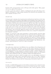
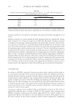
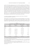
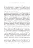
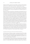
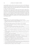
JOURNAL OF COSMETIC SCIENCE 542 Exposure to light alone did not signifi cantly reduce the survival of fi broblasts. Exposure to TiO2 (anatase) prior to irradiation resulted in a reduction of survival that was depen- dent on the dose of light. The relationship between level of exposure to TiO2 (anatase) and survival is shown in Figure 3. TiO2 (anatase) was not cytotoxic, i.e., no toxicity was seen when fi broblasts were exposed to TiO2 (anatase) alone. However, TiO2 (anatase) was pho- tocytotoxic. Exposure to both TiO2 (anatase) and a combination of 10 J/cm2 UVA radia- tion and 45 J/cm2 visible light resulted in a reduction in survival that was dependent on the level of exposure to TiO2 (anatase). Using the data shown in Figure 3, the PD50 for TiO2 (anatase) was found to be 0.82 ± 0.14 μg/cm2 (Table II). The photocytotoxicity of two samples of commercially available TiO2 (rutile) was similarly examined. One sample of TiO2 (rutile), found by X-ray diffraction to contain entirely the rutile crystalline form, did not elicit photocytotoxicity (Table II). The second sample of TiO2 (rutile), which contained 4.7% TiO2 (anatase), was photocytotoxic (PD50 = 1.2 ± 0.18 Table II). These results demonstrate that TiO2 containing only a small proportion of the anatase crystal- line form can be signifi cantly photocytotoxic. The results of the photocytotoxicity tests for ten permanent makeup inks are shown in Table II. None of the inks were cytotoxic, i.e., the survival of fi broblasts was unaffected Figure 2. UVA dependence for photocytotoxicity elicited by TiO2. Human dermal fi broblasts were incu- bated 18 hours with medium containing 2 μg/cm2 TiO2 (anatase) (open circles). Control samples received no TiO2 (closed circles). Fibroblasts were then irradiated with a combination of UVA radiation and visible light. Survival was assessed by determining the ability of fi broblasts to form colonies following irradiation. Data points represent the average ± standard deviation of four replicates. Figure 3. Concentration dependence for photocytotoxicity elicited by TiO2. Human dermal fi broblasts were incubated 18 hours with medium containing the indicated levels of TiO2 (anatase). Fibroblasts were then irradiated with a combination of 10 J/cm2 UVA radiation and 45 J/cm2 visible light (open circles). Control groups were treated with only TiO2 (anatase) (closed circles). Survival was assessed by determining the abil- ity of fi broblasts to form colonies following irradiation. Data points represent the average ± standard devia- tion of four replicates.
Purchased for the exclusive use of nofirst nolast (unknown)
From: SCC Media Library & Resource Center (library.scconline.org)