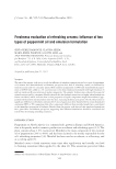
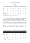
PHOTOCYTOTOXICITY OF TITANIUM DIOXIDE 543 by treatment with the inks alone (data not shown). However, eight inks were found to be photocytotoxic. Six of the photocytotoxic inks (inks 1, 2, 3, 6, 7, and 10) contained pre- dominately TiO2 (anatase). Ink 5 contained comparable amounts of TiO2 (anatase) and TiO2 (rutile). These results indicate that the photocytotoxicity of the inks is attributable to TiO2 (anatase). Ink 4, which contained only 0.6 % TiO2 (anatase), was also determined to be phototoxic. However, ink 4 contained additional pigments as evident from its color (fl esh-colored). These additional, unidentifi ed pigments may have contributed to the photocytotoxicity elicited by ink 4. The remaining inks, which contained only TiO2 (rutile) (inks 8 and 9), were not photocytotoxic. Inks used for permanent makeup are complex mixtures, generally containing mixtures of pigments dispersed in a multicomponent diluent (1). While it is plausible that the pig- ments contained in inks elicited the photocytotoxicity we observed, it is possible that components in the diluent may have enhanced or moderated the photocytotoxic response. To investigate this possibility, pigments were isolated from the inks and tested indepen- dently using the described in vitro method for assessing cytotoxicity and photocytotoxic- ity. The results are presented in Table II. No cytotoxicity was noted following treatment with pigments alone (data not shown). However, every pigment isolated from a photocy- totoxic ink was also photocytotoxic. In addition, pigments isolated from inks that were not photocytotoxic were also not photocytotoxic. These results indicate that the observed photocytotoxicity is attributable primarily to the pigments contained in the inks. For inks 1, 2, and 4, the PD50 determined for the ink was signifi cantly larger than the PD50 determined for the pigments isolated from the inks (Table II). This result suggests that these inks contain components that can moderate the photocytotoxicity of the pigments. We are currently developing methods to fractionate permanent makeup inks to deter- mine how interactions between components of an ink affect the ink’s photocytotoxicity. In addition to crystalline form, particle size and surface coating can moderate the photo- catalytic activity of TiO2. The reactions photocatalyzed by TiO2, and resulting in the formation of ROS, occur at the particle’s surface. Because the surface-area-to-mass ratio decreases as particle size increases, TiO2 composed of large particles has diminished pho- tocatalytic catalytic activity due to the relatively lower surface area. In addition, applica- tion of a surface coating is a common strategy for moderating the photocatalytic activity of TiO2 (24). Frequently used surface coatings include inorganic oxides (e.g., Al2O3 and SiO2), dimethicone, and stearic acid (40,41). Analysis by X-ray fl uorescence suggests that some of the inks (inks 2, 4, 8, 9, and 10) are formulated using TiO2 coated with Al2O3 or a combination of Al2O3 and SiO2 (Table I). However, several of these inks (inks 2, 4, and 10) were found to be photocytotoxic (Table II). These results are consistent with reports that coating TiO2, particularly the anatase crystalline form, with inorganic oxides pro- vides incomplete protection against photocatalysis and photocytotoxicity (36,40,42). GENERATION OF THE HYDROXYL RADICAL FOLLOWING PHOTOEXCITTION OF PIGMENTS ISOLATED FROM PERMANENT MAKEUP INKS Investigators have shown that ESR is a useful method for detecting ROS formed during photoexcitation of TiO2. The mechanisms underlying the photocatalytic activity, and particularly the photocytotoxicity, of TiO2 are not well understood. However, available studies indicate that the hydroxyl radical is the primary ROS responsible for the chemical and biochemical events initiated by photoexcited TiO2 (31,43). Therefore, we have mea- sured the formation of the hydroxyl radical to characterize the ROS generated during

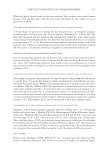
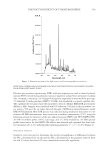
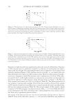
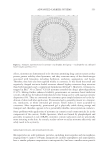
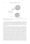
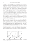
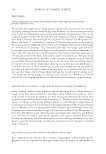
JOURNAL OF COSMETIC SCIENCE 544 photoexcitation of pigments isolated from permanent makeup inks. Figure 4 shows the effect of irradiation time on the ESR signal for a suspension containing the pigment iso- lated from ink 6, which was shown by X-ray diffraction to contain 100% TiO2 (anatase) (Table II). A four-line ESR signal, characteristic of the spin adduct between DMPO and the hydroxyl radical (DMPO-OH), was observed. The signal intensity increased with increasing irradiation times up to 20 minutes (Figure 4). Figure 5 shows the relationship between the concentration of the pigment from ink 6 and the intensity of the ESR signal, measured as the peak-to-peak height of the second line in the ESR spectrum for DMPO-OH. All samples were irradiated at 320 nm. The intensity of the ESR signal in- creased for suspensions containing up to 50 μg/ml of pigment and began to plateau thereafter. These results established the appropriate irradiation time (20 min) and con- centration (50 μg/ml) for measuring effi ciencies of hydroxyl radical generation. The effi ciencies for hydroxyl radical generation, evaluated as the ESR intensity of the sample relative to the ESR intensity of commercially available TiO2 (anatase), are shown in Table II. Except for the pigment isolated from ink 10, all pigments containing TiO2 (anatase) photocatalyzed the formation of hydroxyl radicals. There did not appear to be a correlation between the percentage of TiO2 (anatase) and the effi ciency of hydroxyl radical formation for pigments isolated from inks. This suggests that other properties, such as surface coating and particle size, signifi cantly infl uence the effi ciency of these pigments to photocatalyze the formation of hydroxyl radicals. Similarly, except for the pigment isolated from ink 10, all pigments that elicited photocytotoxicity also photocatalyzed the formation of hydroxyl radicals. These results are consistent with the role of ROS in the photocytotoxicity elicited by TiO2. However, there was little correlation between the PD50 measured for a pigment and its effi ciency for photocatalyzing the formation of the Figure 4. UVA dependence for the formation of the adduct between the hydroxyl radical and the spin trap, DMPO. The pigment isolated from ink 6 (50 μg/ml), suspended in water containing 50 mM DMPO, was irradiated for the indicated times with UV radiation (320 nm). The spin adduct, DMPO-OH, was detected by ESR as described in the Experimental section.
Purchased for the exclusive use of nofirst nolast (unknown)
From: SCC Media Library & Resource Center (library.scconline.org)