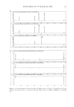
ANTIBIOTIC SUSCEPTIBILITY AND UVc LIGHT 137 DNA, thus leading to cell death. UVc light has a wavelength of between 280 and 200 nm and has the highest energy, compared to UVa and UVb light. Due to its absorption in the lithosphere, relatively little UVc light is experienced on the earth’s surface, in compari- son to UVa and UVb light, with the exception of high altitudes. Equally, cosmetic UV sunbeds do not emit UV light within the UVc range. Therefore, it would appear an usual choice of light on which to perform these experiments. UVc radiation was selected due to its highest energy and thus its ability to infl ict the greatest ability for bacterial mutational change and genomic re-organization, including alterations to the organism’s antibiotic susceptibility. Staphylococcal organisms were exposed to UVc radiation until the point of lethality, and their antibiotic susceptibility was examined along this time course. Very little is known about the effects of UV sunbeds on altering the diversity of the host bacterial commensal skin fl ora. Any UV radiation-induced alteration may led to a bacte- rial ecological imbalance, thereby leading to a vulnerability for the colonization and sub- sequent infection with skin pathogenic organisms. Overall, we were not able demonstrate any alteration in antibiotic resistance levels in any of the staphylococcal skin organisms examined, suggesting that UV stress response is not linked with upregulation of a global stress response cascade within the organisms examined. In conclusion, these in vitro data do not support any alteration in antibiotic susceptibility when challenged with sublethal stress from UVc light. ACKNOWLEDGMENT This work was fi nancially supported through an HSC R&D Offi ce commissioned grant: Antimicrobial Resistance Action Plan (AMRAP) (COM/2730/04). REFERENCES (1) D. E. Fisher and W. D. James, Indoor tanning—Science, behavior, and policy, New Eng. J. Med., 363, 901–903 (2010). (2) M. A. McMahon, J. Xu, J. E. Moore, I. S. Blair, and D. A. McDowell, Environmental stress and antibi- otic resistance in food-related pathogens, Appl. Environ. Microbiol., 73, 211–217 (2007). (3) P. Gilbert and A. J. McBain, Potential impact of increased use of biocides in consumer products on prevalence of antibiotic resistance, Clin. Microbiol. Rev., 16, 189–208 (2003). (4) A. P. Schuch and C. F. Menck, The genotoxic effects of DNA lesions induced by artifi cial UV-radiation and sunlight, J. Photochem. Photobiol. B, 99, 111–116 (2010). (5) Clinical and Laboratory Standards Institute (CLSI), Performance standards for antimicrobial susceptibil- ity testing. Document M100-S15 (Wayne, PA, 2005). (6) M. N. Alekshun and S. B. Levy, Alteration of the repressor activity of MarR, the negative regulator of the Escherichia coli marRAB locus, by multiple chemicals in vitro, J. Bacteriol., 181, 4669–4672 (1999). (7) A. H. Rickard, S. Lindsay, G. B. Lockwood, and P. Gilbert, Induction of the mar operon by miscella- neous groceries, J. Appl. Microbiol., 97, 1063–1068 (2004). (8) N. J. Rowan, Evidence that inimical food-preservation barriers alter microbial resistance, cell morphol- ogy and virulence, Trends Food Sci. Technol., 10, 261–270 (1999). (9) J. Marles-Wright, T. Grant, O. Delumeau, G. van Duinen, S. J. Firbank, P. J. Lewis, J. W. Murray, J. A. Newman, M. B. Quin, P. R. Race, A. Rohou, W. Tichelaar, M. van Heel, and R. J. Lewis, Molecular architecture of the “stressosome,” a signal integration and transduction hub, Science, 322(5898), 92–96 (2008).

Purchased for the exclusive use of nofirst nolast (unknown)
From: SCC Media Library & Resource Center (library.scconline.org)