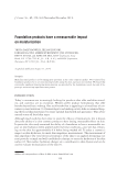
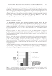
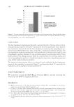
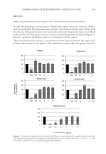
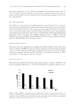
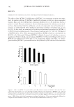
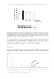
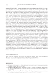
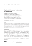
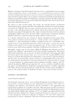
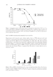
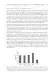
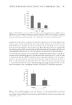
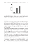
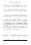
NELUMBO NUCIFERA AND INHIBITED TYROSINASE ACTIVITY AND MELANOGENESIS 383 without cytotoxicity. Indicated concentrations of JKTM-12 (10, 50, and 100 μg/ml) were pretreated in B16F10 cells for 1 h, and then 100 nM α-MSH was added to the cells to stimulate melanin biosynthesis in the cells. After 48 h, melanin level was detected using melanin assay described in the experimental section. Melanin was increased by 100 nM α-MSH treatment in B16F10 cells however, pretreatment of 50 μg/ml and 100 μg/ml JKTM-12 inhibited the melanin level (Figure 3B). Moreover, 100 μg/ml JKTM-12 in- hibited the cellular tyrosinase activity and tyrosinase protein expression by western blot- ting (Figure 6). These result demonstrated that JKTM-12 inhibits melanin biosynthesis without cell cytotoxicity. INHIBITION OF MELANIN BIOSYNTHESIS AND TRP-1, TRP-2 mRNA EXPRESSION BY HYPEROSIDE AND ASTRAGALIN We tested tyrosinase activity using the components of JKTM-12 ethanol extract. Inter- estingly, the receptables of N. nucifera inhibited tyrosinase activity more strongly than the Figure 4. Effects of hyperoside and astragalin on (A) viability and (B) melanin contents in B16F10 cells. Cells were cultured with the indicated concentrations of hyperoside and astragalin and then processed for the analysis of viability and melanin contents. Data are presented as the mean ± SEM of three individual experi- ments, performed in duplicate. *p 0.05 versus the only α-MSH-treated control values.

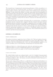
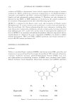
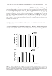
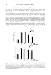
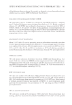
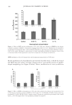
JOURNAL OF COSMETIC SCIENCE 384 fl owers, roots, and seeds of N. nucifera (data not shown). In our previous study, we isolated two bio-active compounds (hyperoside and astragalin) from an n-EtOAc fraction using the receptacles of N. nucifera. Hyperoside (IC50 = 15.67 μg/ml) and astragalin (IC50 = 21.22 μg/ml) were shown to inhibit tyrosinase activity (13). In this study, we demon- strated that hyperoside and astragalin inhibit melanine synthesis, TRP-1 mRNA, TRP-2 mRNA expression, cellular tyrosinase activity, and tyrosinase protein level. Hyperoside and astragalin were not cytotoxic to melanoma cells at the indicated concentrations (Fig- ure 4A). However, pretreatment of hyperoside and astragalin suppressed melanin biosyn- thesis overexpressed by α-MSH in B16F10 cells in a dose-dependent manner (Figure 4B). Because TRP-1 and TRP-2 are known as melanogenic enzymes, we investigated whether hyperoside and astragalin regulated TRP-1 and TRP-2 mRNA expression. As shown in Figure 5, TRP-1 and TRP-2 mRNA were overexpressed by 100 nM α-MSH in 48 h. However, pretreatment of hyperoside and astragalin inhibited TRP-1 and TRP-2 mRNA expression. Cellular tyrosinase and tyrosinase protein level were activated by 100 nM α-MSH, but pretreatment of hyperoside (10 μg/ml) and astragalin (10 μg/ml) inhibited Figure 5. Effects of hyperoside and astragalin on the mRNA expresstion of (A) TRP-1 and (B) TRP-2 in B16F10 cells. Cells were incubated in 6 well plates (2 × 10⁵ cells/well) with the indicated concentrations of hyperoside or astragalin for 1 h and then exposed to 100 nM α-MSH for 48 h. mRNA expression of TRP-1 and TRP-2 was detected by real-time PCR. Hprt1 was used as a loading control. Data are presented as the mean ± SEM of three individual experiments, performed in triplicate. *p 0.05 and **p 0.01 versus the only α-MSH-treated control values.
Purchased for the exclusive use of nofirst nolast (unknown)
From: SCC Media Library & Resource Center (library.scconline.org)