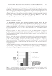
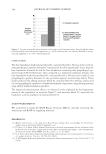
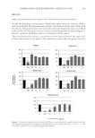
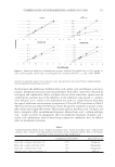
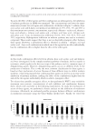
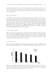
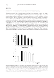
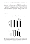
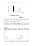
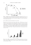
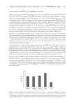
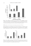
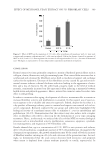
EFFECT OF METHANOL YEAST EXTRACT ON 3T3 FIBROBLAST CELLS 391 cell proliferation (data not shown). As a result, we obtained a viscous brownish and water soluble bioactive complex (MYE for Methanol Yeast Extract). EVALUATION OF THE ANTIOXIDANT PROPERTY OF MYE The antioxidant capacity of MYE was evaluated by the DPPH method (1,1 diphenyl pycril 2 hydrazil) following a protocol adapted from Szabo et al. (21). Briefl y, a methanol solution of 1 mM DPPH was incubated in the presence or absence (control) of MYE at 0.001%, 0.01%, 0.1%, and 1% v/v. After 15 min incubation at room temperature, ab- sorbance at 517 nm was measured using an Oasis UVM340 spectrophotometer. Antioxi- dant effect of the yeast extract was compared to the one of ascorbic acid at concentrations ranging from 0.001 to 1mM. MEASUREMENT OF YEAST PROLIFERATION About 5 × 10⁶ cells of S. cerevisiae previously grown on rich medium were used to inoculate 50 ml of glycerol minimal medium (4% glycerol/0.17% yeast nitrogen base/0.5% ammo- nium sulphate Difco, Detroit, MI) (22) supplemented or not (control) with MYE at 0.01%, 0.1%, and 1%. After 24, 48, and 96 h of incubation at 30°C with shaking, growth was assessed by measuring the absorbance at 660 nm using a Unicam UV1 spectrophotometer. ANIMAL CELL CULTURE 3T3 cells (mouse embryonic fi broblasts) were from DSMZ bank (Braunschweig, Ger- many). Cells were cultured in DMEM medium supplemented with 4 mM glutamine, 100 U/ml penicillin, 100 μg/ml streptomycin and 10% heat-inactivated fetal bovine se- rum (FBS) (all reagents from Invitrogen/Gibco, Carlsbad, CA). Cells were maintained at 37°C in a humid atmosphere containing 5% CO2. MTT CYTOTOXICITY ASSAY 3T3 cells were seeded in 96-well plates (3000 cells/well). Twenty four hours after seed- ing, cells were treated or not (control) with MYE at 0.01%, 0.1%, and 1% for a period of 48 h. Cell viability was then assayed by exposure to 0.03% MTT w/v. After removal of medium, produced formazan was dissolved in DMSO (1 h, room temperature, under agitation) for measurement by spectrometry at 550 nm using an Oasis UVM340 spectrophotometer. 3T3 CELL GROWTH MEASUREMENT 3T3 cells were seeded in 96-well plates (3000 cells/well). Twenty four hours after seeding, cells were treated or not (control) with MYE at 0.01% and 0.1% for a period

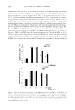
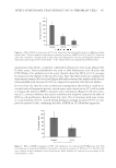
JOURNAL OF COSMETIC SCIENCE 392 of 72 h. Cell growth was then measured by crystal violet staining as previously de- scribed (23). Briefl y, cells were washed with PBS, fi xed for 15 min in glutaraldehyde (1% in PBS), and stained for 30 min with crystal violet (0.1% w/v in distilled water). After removal of dye excess, cell-bound crystal violet was extracted with 1% v/v Triton X-100 and absorbance was measured at 550 nm using an Oasis UVM340 spectrophotometer. CLONOGENICITY STUDY 3T3 cells were seeded in 6-well dishes at low concentration (100 cells/well). Twenty four hours after seeding, cells were treated or not (control) with MYE at 0.01% and 0.1% for 72 h. Cells were then fi xed and stained with crystal violet as described above. After staining, visible clones (at least about 20 cells) were counted and diameter of the 10 largest colonies was recorded. SENESCENCE ASSAY 3T3 cells were seeded in 6-well plates (105 cells/well) and incubated for 96 h in DMEM containing 2% FBS. Cells were then treated or not (control) with MYE at 0.01% and 0.1% and incubated for an additional 72 h in DMEM containing 2% FBS. Senescence was evaluated via the detection of β-galactosidase activity using the Senescence Detection Kit from Merck Biosciences (Schwalbach, Germany) according to the manufacturer’s instructions. TUNEL ASSAY FOR APOPTOSIS DETECTION 3T3 cells were seeded in 6-well plates (105 cells/well) and allowed to grow for 24 h. Me- dium was then replaced by serum-free DMEM containing or not (control) MYE at 0.1% for 48 h. Fragmented DNA was then biotinilated by using the DeadEnd tunnel assay system from Promega (Madison, WI), labeled with streptavidin-coupled Dylight 594 (Invitrogen) and visualized by fl uorescence microscopy. ACTIN CYTOSKELETON REARRANGEMENT STUDY 3T3 cells were seeded in 8-chamber Lab-Tek slides and allowed to grow for 48 h before treatment with or without (control) MYE at 0.1%. After 90 min of incubation, monolay- ers were washed twice with PBS, fi xed in a 3% paraformaldehyde–PBS solution (20 min, room temperature), permeabilized with a 0.1% Triton X-100-PBS solution (5 min, room temperature, under agitation), and nonspecifi c binding side were blocked with a 2% BSA–PBS solution (overnight, 4°C). Cells were then exposed for 1 h to 1 μg/ml Texas red-labeled phalloidin (for F-actin) or 10 μg/ml FITC-labeled DNAse I (for G-actin), both diluted in a 2% BSA–PBS solution. After several washes with a 0.05% Tween 20- PBS solution, F- and G-actin were visualized by fl uorescence microscopy. In addition,
Purchased for the exclusive use of nofirst nolast (unknown)
From: SCC Media Library & Resource Center (library.scconline.org)