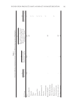
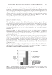
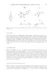
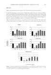
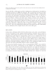
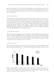
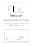
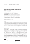
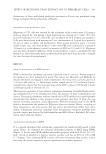
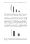
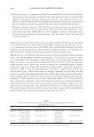
COMBINATION OF DEPIGMENTING AGENTS IN VITRO 369 RESULTS EFFECT OF ARBUTIN, KOJIC ACID, AZELAIC ACID, AND α-LIPOIC ACID ON CELL VIABILITY To fi nd the maximum concentration at which these agents were not cytotoxic, WST-1 assay was performed. The maximum non-cytotoxic concentrations obtained were 1000 μg/ml for arbutin, 100 μg/ml for kojic acid, and azelaic acid and 10 μg/ml for lipoic acid, which indicates that the four agents are less cytotoxic than hydroquinone (5 μg/ml) (Figure 2). Besides, signifi cant proliferative effect was observed in all the agents. These maximum non-cytotoxic concentrations were used to perform the same assay doing combinations of two agents. This was done to ensure that the agents, used in a Figure 2. Human skin melanocytes viability with different concentrations of the agents cultured individu- ally. Results are expressed as percentage of cell viability relative to control. Values of p 0.05 (*), p 0.01 (**), and p 0.001 (***) are considered signifi cantly different.

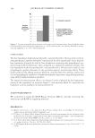
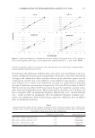
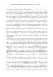
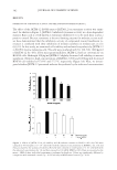
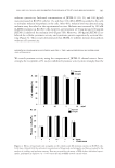
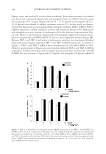
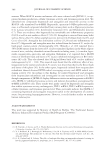
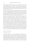
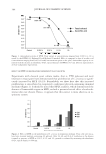
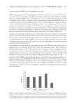
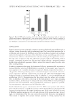
JOURNAL OF COSMETIC SCIENCE 370 non-cytotoxic concentration, were still not cytotoxic in combination with others. As shown in Figure 3, only arbutin + α-lipoic acid combination had a signifi cantly cytotoxic effect on HSM proliferation at their maximum non-cytotoxic concentration individually. Besides, as it happened with the individual treatment, arbutin + azelaic acid, arbutin + kojic acid, and arbutin + α-lipoic acid combinations showed a signifi cant proliferative effect on HSM cells. ARBUTIN, KOJIC ACID, AZELAIC ACID, AND α-LIPOIC ACID MUSHROOM TYROSINASE KINETIC ANALYSIS To fi nd the type of inhibition of these agents over mushroom tyrosinase, different concentra- tions of substrate (L-DOPA) in the presence of the inhibitors were used. Low concentrations of inhibitor were used to see possible synergistic effects between them. Lineweaver–Burk plots of 1/v versus 1/[L-DOPA] were made and showed that arbutin, kojic acid, α-lipoic acid, and azelaic acid are competitive, mixed, competitive, and competitive inhibitors, re- spectively, on diphenolase activity of mushroom tyrosinase (Figure 4). Figure 3. Human skin melanocytes viability with the maximum non-cytotoxic concentrations of the agents combined and its dilutions. Concentrations are expressed as concentration of the fi rst agent/concentration of the second agent. Results are expressed as percentage of cell viability relative to control. Values of p 0.05 (*), p 0.01 (**), and p 0.001 (***) are considered signifi cantly different.
Purchased for the exclusive use of nofirst nolast (unknown)
From: SCC Media Library & Resource Center (library.scconline.org)