JOURNAL OF COSMETIC SCIENCE 392 of 72 h. Cell growth was then measured by crystal violet staining as previously de- scribed (23). Briefl y, cells were washed with PBS, fi xed for 15 min in glutaraldehyde (1% in PBS), and stained for 30 min with crystal violet (0.1% w/v in distilled water). After removal of dye excess, cell-bound crystal violet was extracted with 1% v/v Triton X-100 and absorbance was measured at 550 nm using an Oasis UVM340 spectrophotometer. CLONOGENICITY STUDY 3T3 cells were seeded in 6-well dishes at low concentration (100 cells/well). Twenty four hours after seeding, cells were treated or not (control) with MYE at 0.01% and 0.1% for 72 h. Cells were then fi xed and stained with crystal violet as described above. After staining, visible clones (at least about 20 cells) were counted and diameter of the 10 largest colonies was recorded. SENESCENCE ASSAY 3T3 cells were seeded in 6-well plates (105 cells/well) and incubated for 96 h in DMEM containing 2% FBS. Cells were then treated or not (control) with MYE at 0.01% and 0.1% and incubated for an additional 72 h in DMEM containing 2% FBS. Senescence was evaluated via the detection of β-galactosidase activity using the Senescence Detection Kit from Merck Biosciences (Schwalbach, Germany) according to the manufacturer’s instructions. TUNEL ASSAY FOR APOPTOSIS DETECTION 3T3 cells were seeded in 6-well plates (105 cells/well) and allowed to grow for 24 h. Me- dium was then replaced by serum-free DMEM containing or not (control) MYE at 0.1% for 48 h. Fragmented DNA was then biotinilated by using the DeadEnd tunnel assay system from Promega (Madison, WI), labeled with streptavidin-coupled Dylight 594 (Invitrogen) and visualized by fl uorescence microscopy. ACTIN CYTOSKELETON REARRANGEMENT STUDY 3T3 cells were seeded in 8-chamber Lab-Tek slides and allowed to grow for 48 h before treatment with or without (control) MYE at 0.1%. After 90 min of incubation, monolay- ers were washed twice with PBS, fi xed in a 3% paraformaldehyde–PBS solution (20 min, room temperature), permeabilized with a 0.1% Triton X-100-PBS solution (5 min, room temperature, under agitation), and nonspecifi c binding side were blocked with a 2% BSA–PBS solution (overnight, 4°C). Cells were then exposed for 1 h to 1 μg/ml Texas red-labeled phalloidin (for F-actin) or 10 μg/ml FITC-labeled DNAse I (for G-actin), both diluted in a 2% BSA–PBS solution. After several washes with a 0.05% Tween 20- PBS solution, F- and G-actin were visualized by fl uorescence microscopy. In addition,

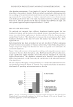
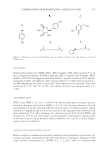
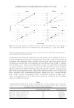
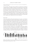
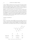
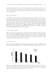
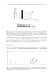
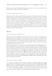
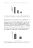
EFFECT OF METHANOL YEAST EXTRACT ON 3T3 FIBROBLAST CELLS 393 fl uorescence of Texas red-labeled phalloidin associated to F-actin was quantifi ed using Image J software (National Institute of Health). MIGRATION STUDY BY SCRATCH ASSAY Migration of 3T3 cells was assessed by the technique of the scratch assay (24) using a protocol adapted for cells having a high migration rate (wound of 2.2 mm). 3T3 cells, seeded 6-well plates (5 × 10⁵ cells/well), were incubated for 48 h without any treatment. Cells were then treated with mitomycin C at a concentration of 5 μg/ml for a period of 90 min in order to inhibit cell proliferation. After wound formation using a 2.2-mm width scraper (t0), cells were washed 3 times with PBS and incubated for a period of 24 hours (t24) in the absence (control) or presence of MYE at 0.01 and 0.1%. Migration rate was then calculated (difference of the wound width at t0 and t24 normalized by the width at t0). Note that marks made on the back of the plate were done in order to identify the exact location of measurements. RESULTS MAIN CHARACTERISTICS OF MYE EXTRACT MYE is obtained by methanol extraction of pressed yeast S. cerevisiae. Various stages of precipitation are then performed to purify this extract (see Materials and Methods for details). Brownish and liquid at room temperature, this extract has a specifi c gravity of 1.41 and a pH value of 7.5. Preliminary studies carried out by G-25 exclusion chroma- tography have shown that the molecular mass of the MYE is about 1500 Da and it shows an absorption peak at 260 nm (16,17). The bioactive properties of yeast extracts containing chromium or soluble β-glucans be- ing established (9–15), we attempted to detect the presence of these compounds in our extract. Concerning chromium, analysis carried out by ICP (inductively coupled plasma) showed that this element is only present in trace amounts (less than 10 ppm data not shown). Similarly, enzymatic assays (Megazyme, Wicklow, Ireland) made on the MYE failed to demonstrate the presence of β-glucans (data not shown). Hence, according to these experiments, one may assume that the extract described here differs from chro- mium- or β-glucans-rich yeast complexes. Note that presence of amines, probably from peptidic origin, was detected by both ninhydrin and BCA (Pierce, Rockford, IL) colori- metric assays (data not shown). ANTIOXIDANT PROPERTIES OF MYE It is a general agreement that antioxidant compounds are benefi cial with regard to skin aging. Hence, we evaluated the potential antioxidant property of MYE using DPPH (Figure 1). In this assay, MYE exhibits antioxidant property with an IC50 of 1.16 × 10³ ppm. Tested in parallel as a positive control, ascorbic acid has an IC50 of 0.034 mM (= 5.9 ppm). Thus, although MYE is about 200 times less potent than ascorbic acid in this assay, this extract appears to possess signifi cant antioxidant properties.
Purchased for the exclusive use of nofirst nolast (unknown)
From: SCC Media Library & Resource Center (library.scconline.org)