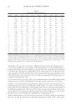
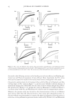
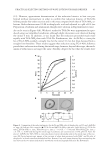
JOURNAL OF COSMETIC SCIENCE 4 30 min at room temperature. Subsequently, the absorbance of the mixture was measured using the UV/VIS spectrophotometer (OPTIZEN 2020UV plus, Mecasys Co., Daejeon, Korea) at a wavelength of 750 nm. Using Tannic acid (Sigma-Aldrich Korea Ltd, Youngjin-city, Korea) as a standard substance, the replicates were tested three times for the analysis. Flavonoid measurement. The fl avonoids content per gram of extract was measured using the colorimetric method in the presence of diethylene glycol (13). Approximately 100 μl of the extract, 100 μl of 1 N NaOH, and 1 ml of diethylene glycol were combined using a vortex mixer and incubated for 1 h in a 30°C water bath. Following the reaction, the absorbance of the mixture was measured using a UV/VIS spectrophotometer at a wave- length of 420 nm. Catechin (Sigma-Aldrich Korea Ltd) was used as a standard substance for three repeat examinations. DPPH radical scavenging ac tivity measurement. The DPPH radical scavenging activity was measured using a modifi ed version of the method described by Blois (14). First, we pro- duced 500 mM of DPPH using 0.1 M Tris base-HCl buffer (Tris buffer) at pH 7.4 and methanol. Then, a sample aliquot of 100 μl, 400 μl of 0.1 M Tris buffer and 500 μl of 500 mM DPPH were combined and vortexed. The mixture was reacted for 30 min in the dark at room temperature and the absorbance of the mixture was analyzed using a UV/VIS spec- trometer at 517 nm in three replicate runs. The artifi cial antioxidant butylated hydroxytoluene (BHT) was used as a control and the DPPH radical scavenging activity was expressed as electron donation abilit y (EDA) percent- age, a percentage based on the differences in absorbance of DPPH solutions between absence (A_initial) and presence (A_fi nal) of antioxidant samples (BHT, MLFE, or MEFE) (15): EDA (%) = (A_initial – A_fi nal/A_initial) × 100%. Cell viability estimation using MTT assay. Cell viability was measured using the methodol- ogy of Mosmann (16). RAW 264.7 cells were seeded onto 96-well plates at a density of 3 × 10⁵ cells/well and cultured at 37°C in a 5% CO2 incubator. The cells were cultured for another 24 h after treatment using various concentrations of the MLFE and MEFE extracts. After culturing, 100 μl of MTT solution was placed into each well, cultured for 4 hours, and the formazan precipitate was then dissolved in 100 μl of dimethylsulfoxide after the removal of the supernatant. An ELISA microplate reader (E-max, Molecular Device, Sunnyvale, CA) was used to measure the absorbance at 570 nm in three replicate viability assays. Cell viability is expressed in percentage using the following formula (17): Cell viability (%) = (A−B)/A × 100% A: Absorbance of untreated RAW 264.7 cells at 570 nm B: Absorbance of extract sample at 570 nm. ROS production measurement. RAW 264.7 cells (4 × 10⁵ cells) were seeded in a glass bottom dish (MatTek Corp., Ashland, MA) and incubated for 24 h. Cells were treated with a various amounts of the MLFE or MEFE for 10 hours and incubated for 20 h with 1 ml of 1 μg/ml LPS (Sigma-Aldrich Korea Ltd: E. Coli 0111:B4, L3024). Then, cells were treated with 10 μM of dichlorofl uorescein-diacetate (DCFH-DA, Sigma-Aldrich) for 0.5 h and harvested with trypsin/EDTA (Gibco) for three separate fl ow cytometric analyses.

ANTIOXIDANT PROPERTIES OF FERMENTED MANGO LEAF EXTRACTS 5 DCFH-DA is a non-fl uorescent material that can permeate cell membranes. Within cells, it hydrolyzes to dichlorodihydrofl uorescein (DCFH), which in the presence of ROS is oxi- dized to fl uorescent dichlorofl uorescein (DCF), a high-performance fl uorescent substance. To quantify the fl uorescent cells, fl ow cytometry was also performed using a Flow Cytom- etry Caliber (Becton Dickinson, Franklin Lakes, NJ) with CellQuest Pro software. The percentage of cells in positive events was calculated as the events within the gate divided by total number of events then subtracting percentage of the control sample (untreated cells) (18). Tyrosinase inhibitory activity estimation. Tyrosinase inhibition activity was assessed using the modifi ed protocol of Masamoto et al. (19). To det ermine in vitro mushroom tyrosinase inhibition activity, 0.3 ml of 2.5 mM 3,4 dihydroxyphenylalanine (L-DOPA), 0.05 ml of the mango extract, and 1.5 ml of 0.1 M phosphate buffer solution (pH 6.8) were com- bined using a vortex mixer and pre-incubated at 25°C. Appro ximately 0.05 ml of mushroom tyrosinase (1.380 units/ml, Sigma-Aldrich) was placed into the vortex mixer and allowed to react for 2 min at 25°C. The absorbance was then measured using a UV/VIS spectrophotometer at a wavelength of 475 nm. The fol- lowing equation was used in the calculations for the percentage of tyrosinase inhibition: Tyrosinase inhibition (%) = [(A−B)/A] × 100% A = Difference in absorbance between 0.5 and 1 minutes in the reactant without the sample B = Difference in absorbance between 0.5 and 1 minutes in the reactant with the sample using 0.05 ml of 1380 units/ml mushroom tyrosinase. Nitrite-scavenging ability assessment. Nitrite scavenging was assessed using a modifi ed ver- sion of Gray and Dugan’s methodology ( 20). We combined 0.1 ml of 1 mM NaNO2 and 0.3 ml of sample MLFE and MEFE, respectively. Then, 0.2 M citrate buffer-HCl (pH 2.5) was added to produce the fi nal volume of 1 ml. The components were combined using a vortex mixer and incubated in a water bath at 37°C for 1 h, after which Griess’ reagents [30% acetic acid solution dissolved 1% sulfa- nilic acid and 1% naphthylamine = 1:1 (v/v)] and 3 ml of 2% acetic acid solution were added. After incubating for 15 min at room temperature, the absorbance of the mixture was measured using a UV/VIS spectrophotometer at a wavelength of 520 nm in three repeat examinations. The nitrite scavenging activity (%) was calculated using the follow- ing formula (21): Nitrite scavenging (%) = [1−(A−B)/C] × 100% A: Absorbance of test sample at 520 nm B: Absorbance of sample blank with H20 instead of NaNO2 at 520 nm C: Absorbance of control (H20) without test sample at 520 nm. STATISTICAL ANALYSIS This study is conducted to test the null hypothesis of equality of antioxidant activities for the MLFE and MEFE. The antioxidant activities at each concentration were expressed as
Purchased for the exclusive use of nofirst nolast (unknown)
From: SCC Media Library & Resource Center (library.scconline.org)