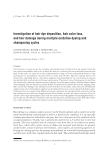
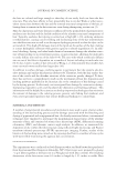
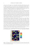
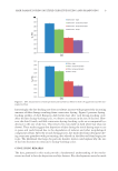
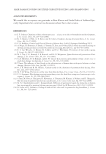
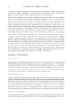
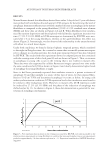
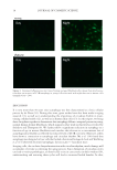
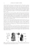
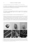
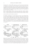
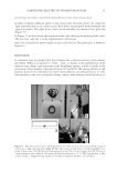
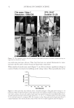
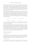
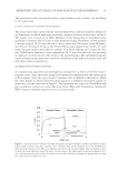
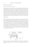
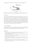
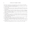
JOURNAL OF COSMETIC SCIENCE 8 AFM (Figure 4) reveals that the fi ber’s internal structure is severely damaged. Cracks (over 100 nm in diameter) and holes are observed in the cuticle (e.g., cell membrane complex, endocuticle) and cortex (e.g., cell membrane complex). Such a result suggests that the damage caused by the bleaching process facilitates dye loss from the internal structure of the hair. To evaluate hair protein damage during the consecutive dyeing–leaching cycles, protein Td of both dyed European white and dark brown hair were measured after each dyeing and leaching cycle. The results are displayed in Figure 5. The fi rst three column bars in each chart demonstrate that Td decreases with consecutive dyeing–leaching cycles, indi- cating that consecutive dyeing cycles progressively degrade hair proteins. More specifi - cally, the amorphous matrix becomes more viscous (or plastic) due to damage to its proteins. It should be noted that Td represents the denaturation temperature of crystalline phase of the hair however, changes to the amorphous matrix (which provides support for the crystalline phase) infl uence the value of Td. Plasticization with water, e.g., shifts Td to lower temperature, while cross-linking hair (making the amorphous matrix more brittle) with reactive ingredients increases Td. The last two column bars in each chart in Figure 5 refl ect how the leaching process alone affects the hair protein structure. We found that hair dyeing alone affects Td more than hair dyeing in combination with leaching. Perhaps the leaching process aids in the recov- ery of hair protein structure. Dyeing the hair results in a large increase in pH to ~9. It requires extensive rinsing to lower the pH to 6.5. It is possible that when conducting DSC measurements at high pH the hair protein is in a highly compromised state. These fi ndings are consistent in both pigmented and nonpigmented hair. Figure 4. AFM images acquired from hair cross sections in (A) virgin hair and (B) bleached then dyed hair. The images in (A) and (B) on the left side are of both cuticle and cortex the images in (A) and (B) in the middle are from the cuticle region and the images in (A) and (B) on the right side are from the cortex region.

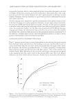
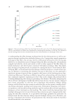
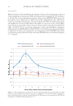
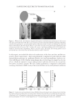
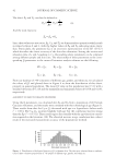
HAIR DAMAGE DURING MULTIPLE OXIDATIVE DYEING AND SHAMPOOING 9 Interestingly, the dye-leaching rate does not always increase with progressively increasing amounts of fi ber damage resulting from consecutive dyeing. Figure 6 presents dyeing– leaching profi les of dyed European dark brown hair after each dyeing–leaching cycle. After the fi rst dyeing–leaching cycle, we observe an increase in the rate of dye loss. How- ever, the third, fourth, and fi fth consecutive dyeing–leaching cycles are accompanied by a decrease in the rate of dye loss. This trend is less noticeable in dyed white hair (data not shown). These results suggest that deposition of dyes during the initial dyeing steps occurs in pores and voids formed due to the degradation of melanin and other morphological components of hair. After the second dyeing process, dye molecules from subsequent dye- ing steps may granulate with pre-existing dyes already in the fi ber and form larger par- ticles. The likelihood that large dye particles dissolve slower could explain why the rate of dye loss decreases on consecutive dyeing–leaching cycles. CONCLUDING REMARKS The data generated in this study provide a fundamental understanding of the mecha- nisms involved in hair dye deposition and dye fastness. Key developments were also made Figure 5. DSC measurements of multiple dyed and leached hair fi bers for both (A) pigmented and (B) non- pigmented hair.
Purchased for the exclusive use of nofirst nolast (unknown)
From: SCC Media Library & Resource Center (library.scconline.org)