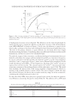
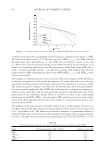
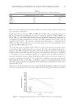
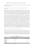
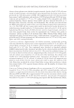
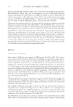
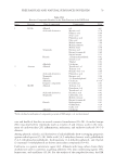
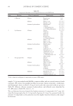
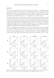
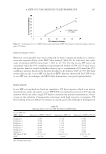
JOURNAL OF COSMETIC SCIENCE 74 version 6.9. Method: (a) mass range from 50 to 800 m/z, (b) trap temperature 150°C, (c) manifold temperature 80°C, and (d) transfer line temperature 247°C injection volume 1 μl, splitting 10%. Specifi c data of the program analysis are provided in Table III. To identify the main organic components, two strategies were followed. First, the spectra of identifi able peaks were compared with commercially available electron mass spectrum libraries such as National Institute of Standards and Technology (NIST) (NIST-Fison, Manchester, UK). In this latter case, spectra with at least 93–97% similarity were chosen. Second, GC-MS analysis was repeated using commercially available standard compounds. 31 P-NMR ANALYSIS OF PHOSPHOLIPIDS: GENERAL PROCEDURE The appropriate peloid (20 g) was extracted with CHCl3/MeOH (2:1 v/v ratio), and the solvent was evaporated under reduced pressure. The crude was extracted with CHCl3/ MeOH/KCl (Folch method) to concentrate phospholipids in the organic phase. A solvent mixture of pyridine and CDCl3 (1.6:1.0 v/v) was prepared under anhydrous conditions. Triphenyl methane was used as internal standard at a concentration of 0.1 mol/l in the aforementioned solvent mixture. Cr(III) acetylacetonate (15 mg) was added as relaxation agent to this standard solution. The sample (100 mg) was dissolved in the solvent solution (0.5 ml). 31 P-NMR spectra were recorded on a Bruker (400 MHz, Milan, Italy) spectrometer, and phospholipids were assigned on the basis of the comparison of the chemical shift with commercially available samples, and when necessary by spiking with predetermined quantities of commercial samples. Typically, the sample was analyzed during 180 min of acquisition time (equivalent to a mean value of 10,000 scans). ANTIOXIDANT ACTIVITY Test systems and culture conditions. L5178Y TK+/− clone (3.7.2C) mouse lymphoma cells were obtained from ATCC (CRL-9518™). Generation time, plating effi ciency, and Table II Extraction Yield of DM and WM Nonpolar Fractionsa Sample Amount of extract (mg) Extraction yield (%) DM-Heptane 20 0.4 DM-Cyclohenane 55 1.1 DM-Diisopropyl ether 50 1.0 WM-Heptane 33 0.7 WM-Cyclohenane 35 0.7 WM-Diisopropyl ether 30 0.6 a The extraction yield refers to 5.0 g of starting material. Table III Set of Data for the GC-MS analysis of DM and WM fractions Temperature Rate (°C/min) Hold (min) Total time (min) 50 – 3 3 280 10 5 31

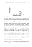
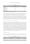
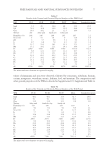
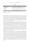
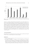
FREE RADICALS AND NATURAL SUBSTANCES IN PELOIDS 75 absence of mycoplasma were checked at regular intervals. Stocks of the L5178Y cells were stored in liquid nitrogen, and subcultures were prepared from the frozen stocks for ex- perimental use. Cells were grown in RPMI 1640 supplemented with 10% heat-inactivated horse serum, 2 mM L -glutamine, and antibiotics (100 IU/ml penicillin and 100 IU/ml strep- tomycin) and incubated at 37°C in a 5% carbon dioxide (CO2) atmosphere and 100% nominal humidity. Chinese hamster ovary (CHO) cells were obtained from Prof. A. T. Natarajan (State University of Leiden, the Netherlands). The cell lines were derived from the CHO isolated from an explant of the ovary of the Chinese hamster (Cricetulus griseus, 2n = 22). The CHO cell line is particularly useful for this kind of studies because of its stable karyotype (modal number is 21 chromosomes), short cell cycle (12–14 h), and its high plating effi ciency. Stocks of CHO cells were stored in liquid nitrogen, and subcul- tures were prepared from these stocks for experimental use. Cultures were grown as monolayer cultures in Ham’s F-10 medium (Gibco BRL, Thermo Fisher, Milan, Italy) supplemented with 15% fetal bovine serum, 4 mM L -glutamine, and antibiotics (50 IU/ml penicillin and 50 IU/ml streptomycin). All incubations were at 37°C in 5% CO2 atmo- sphere and 100% nominal humidity. Cytotoxicity evaluation. Approximately 24 h before treatment, exponentially growing cells were detached by trypsin action, and an appropriate number of 75-cm2 plastic cell culture fl asks containing 15 ml of complete culture medium were individually inocu- lated with 2.0 × 10⁶ cells. Test compounds were dissolved in dimethyl sulfoxide (DMSO) immediately before treatment and added to the culture medium such that the fi nal concentration of solvent did not exceed 1%. The assay was performed using a set of at least six dose levels for each test compound spaced by a factor of 2 (1.0–526 μg/ml), and cell cultures were treated for 3 h. At the end of the treatment, the cultures were washed twice with phosphate-buffered saline, trypsinized, and diluted to obtain an estimated number of 2 × 10³ cells/ml. A volume of 100 μl of each cell suspension was plated in each of three 60-mm tissue culture petri dish to assess the viability of the cells. Plates were incubated for at least 6 days before scoring. After incubation, colonies were stained with a 10% aqueous Giemsa solution, and the number of colonies were scored by hand. Comet assay. Cultures of mouse lymphoma cells at a concentration of 1 × 10⁶ cells/ml were treated for 3 h at 37°C in 5% CO2 atmosphere and 100% nominal humidity, with each sample at a single dose level selected as the dose levels that reduced the relative cloning effi ciency (RCE) of CHO cells to approximately 50% over the concurrent vehicle control values of cultures (Table IV). At the end of the treatment, 10 μl of each cell suspension was added to 65 μl of 0.7% (w/v) low melting point agarose (Bio-Rad Laboratories, Milan, Table IV Dose Levels for Different Fractions Compound Dose level (μg/ml) WM-Acetone 16.0 DM-Acetone 8.0 WM-EtOAc 8.0 DM-EtOAc 8.0 WM-MeOH 128.0 DM-MeOH 64.0
Purchased for the exclusive use of nofirst nolast (unknown)
From: SCC Media Library & Resource Center (library.scconline.org)