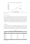
JOURNAL OF COSMETIC SCIENCE 92 (52) S. M. Lynch, B. Frei, Physiological thiol compounds exert pro- and anti-oxidation effects, respectively, on iron- and copper-mediated oxidation of human low-density lipoprotein, Biochem. Biophys. Acta., 1345, 215–221 (1997). (53) L. Mueller, K. Theile, V. Boehm, In vitro antioxidant activity of tocopherols and tocotrienols and com- parison of vitamin E concentration and lipophilic antioxidant capacity in human plasma, Mol. Nutr. Food Res., 54, 731–742 (2010). (54) E. Mocchegiani, L. Costarelli, R. Giacconi, M. Malavolta, A. Basso, F. Piacenza, R. Ostan, E. Cevenini, E. S. Gonos, C. Franceschi, D. Monti, Vitamin E-gene interactions in aging and infl ammatory age- related diseases: Implications for treatment. A systematic review, Ageing Res. Rev., 14, 81–101 (2014). (55) Q. Jiang, Natural forms of vitamin E: Metabolism, antioxidant, and anti-infl ammatory activities and their role in disease prevention and therapy, Free Rad. Biol. Med., 72, 76–90 (2014). (56) N. Duhem, F. Danhier, V. Préat, Vitamin E-based nanomedicines for anti-cancer drug delivery, J. Con- troll. Release, 182, 33–44 (2014). (57) W. Stahl, H. Sies, β-Carotene and other carotenoids in protection from sunlight, Am. J. Clinical Nutr., 96, 1179–1184 (2012). (58) A. T. Dinkova-Kostova, Phytochemicals as protectors against ultraviolet radiation: Versatility of effects and mechanisms, Planta. Med., 74, 1548–1559 (2008). (59) D. Slamenova, E. Horvathova, Cytotoxic, anti-carcinogenic and antioxidant properties of the most fre- quent plant volatiles, Neoplasma, 60, 343–354 (2013). (60) L. Cheng, P. H. Xu, B. D. Shen, G. Shen, J. J. Li, L. Qiu, C. Y. Liu, H, L, Yuan, J. Han, Improve bile duct-targeted drug delivery and therapeutic effi cacy for cholangiocarcinoma by cucurbitacin B loaded phospholipid complex modifi ed with berberine hydrochloride, Int. J. Pharm., 489, 148–157 (2015). (61) E. London, G. W. Feigenson, Phosphorus NMR analysis of phospholipids in detergents, J. Lipid Res., 20, 408–412 (1979). (62) N. W. Lutz, P. J. Cozzone, Multiparametric optimization of 31 P NMR spectroscopic analysis of phos- pholipids in crude tissue extracts. 2. Line width and spectral resolution, Anal. Chem., 82, 5433–5440 (2010). (63) E. Hatzakis, A. Koidis, D. Boskou, P. Dais, Determination of phospholipids in olive oil by 31 P NMR Spectroscopy, J. Agr. Food Chem., 56, 6232–6240 (2008). (64) L. Yao, S. Jung, P-31 NMR phospholipid profi ling of soybean emulsion recovered from aqueous extrac- tion, J. Agr. Food Chem., 58, 4866–4872 (2010). (65) J. Y. Na, K. Song, S. Kim, J. Kwon, Hepatoprotective effect of phosphatidylcholine against carbon tetrachloride liver damage in mice, Biochem. Biophys. Res. Commun., 460, 308–313 (2015). (66) I. S. Kim, M. Y. Hyun, K. Y. Park, C. W. Kim, B. J. Kim, M. N. Kim, Phosphatidylcholine and bipo- lar radiofrequency for treatment of localized fat deposits, J. Dermatol. Treat., 25, 308–309 (2014). (67) S. A. A. Kooijmans, P. Vader, S. M. van Dommelen, W. W. van Solinge, R. M. Schiffelers, Exosome mimetics: A novel class of drug delivery systems, Inter. J. Nanomed., 7, 1525–1541 (2012). (68) Y. H. Lu, J. H. Huang, Y. C. Li, T. T. Ma, P. Sang, W. J. Wang, C. Y. Gao, Variation in nutritional compositions, antioxidant activity and microstructure of Lycopus lucidus Turcz root at different harvest times, Food Chem., 183, 91–100 (2015). (69) N. A. P. Franken, H. M. Rodermond, J. Stap, J. Haveman, C. van Bree, Clonogenic assay of cells in vitro, Nat. Protoc., 1, 2315–2319 (2006). (70) M. Dusinská, Z. Dzupinková, L. Wsólová, V. Harrington, A. R. Collins, Possible involvement of XPA in repair of oxidative DNA damage deduced from analysis of damage, repair and genotype in a human population study, Mutagen, 21, 205–211 (2006).

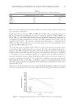
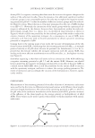
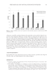
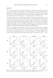
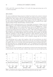
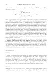
J. Cosmet. Sci., 67, 93–100 (March/April 2016) 93 Use of optical profi lometry and visual grading for measurement of longitudinal striations of the nail CELIA LUDWINSKI, AXELLE CLOCHARD-BOSSUET, THERESA CHEN, KELSEY NORWOOD, and CHRISTIAN ORESAJO, L’Oréal USA Research and Innovation, Clark, NJ 07066. Accepted for publication April 10, 2016. Synopsis Consumer perception of longitudinal striations of the nail is one of the drivers of nail cosmetic purchase and use. The following work investigates the use of objective instrumental methods for the characterization of longitudinal striations. Striations are quantifi ed by Ra (the average maximum height of the profi le), Rq (the root mean square average of the roughness profi le), and Rz (the mean roughness depth), industrial roughness parameters, which are calculated using optical profi lometry of the three-dimensional surface structure of the nail. A visual assessment is conducted by cosmetologists in vivo and on images captured in several lighting conditions. With this evaluation, the cosmetologists provide ratings of surface ridges on a 0–5 scale to com- plement and validate the instrumental method. Both the optical profi lometry and the cosmetologist-graded methods are used to evaluate 33 nails of visually-varying levels of ridges from female volunteers. The evalu- ations from these methods yield well-correlated and repeatable results, and these preliminary fi ndings sug- gest that this new instrumental method can be used to objectively measure longitudinal striations of the nail. INTRODUCTION Consumer perception of cosmetic imperfections of the nail is one of the drivers of nail enamel use. Yellowing, spots, chipping, peeling, thinning, brittleness, and striations are examples of imperfections that can be concealed with cosmetic products. In particular, the appearance of longitudinal striations is a major motivation for the purchase of correc- tive or concealing cosmetic enamels. In light of this trend, this article focuses on the de- velopment of an objective method for the characterization of longitudinal striations. Longitudinal striations can be categorized into two physiological presentations: surface and underface ridging. Surface ridging is what the naive consumer typically refers to as nail ridges and is included in the clinical classifi cation of onychorrhexis. Surface ridging presents as superfi cial parallel ridges and valleys of the nail plate surface, which extend from the proximal nail fold to the distal nail edge (1–5). Comparatively, underface ridging consists of parallel ridges from the proximal nail fold to the distal nail edge Address all correspondence to knorwood@rd.us.loreal.com.
Purchased for the exclusive use of nofirst nolast (unknown)
From: SCC Media Library & Resource Center (library.scconline.org)