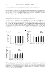
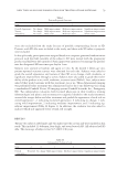
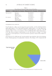
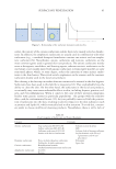
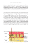
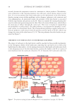
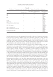
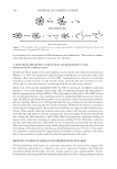
ANTIAGING POTENTIAL OF FUCOXANTHIN 57 MEASUREMENT OF SK IN ELASTICITY Cutometer® MPA 58 0 (Courage + Khazaka GmbH) was used to evaluate changes in skin elasticity at the cheek site. The measurement involved initial deformation of the skin by applying suction at a constant pressure (400–450 mbar). The degree to which the deformed skin returned to its original state was then measured to evaluate the resilience or elasticity of the skin. The skin was suctioned for 2 s at 400 mbar and relaxed for 2 s. The pretension time was set to 0.1 s. In the measurement process, the cutometer MPA580 generated a curve with a distinct shape, from which skin extensibility (Ue), delayed distension (Uv), fi nal deformation (Uf), immediate retraction (Ur), total recovery (Ua), and residual deforma- tion at the end of retraction (R) were obtained. Then, the values for evaluating skin elastic- ity were calculated from the measurement parameters as shown in Table I. Each measurement was repeated three times, and the average value was calculated. EVALUATION OF SKIN WRINKLES USING REPLICAS Silicone solution (Silfl o® Flexico Ltd., Potters Bar, Engl and) was used to make skin replicas. Five drops of the catalyst were dropped on about 5 g of the silicone solution and mixed homogeneously. The mixture was then applied to the periphery of the eye tail, taking care not to generate air bubbles. After about 10 min, when the silicone solution became hard, the replica was removed from the face and stored at room temperature. Wrinkles were evaluated using Visioline® VL650 (Courage + Khazaka GmbH). When the replicas were irradiated with light at an angle, shadows are produced by the fl exion of the wrinkles. Visi- oline® VL650 is a device that captures and analyzes differently generated images depending on the depth of the wrinkles. The roughness of the wrinkles was measured by analyzing high-resolution images using Quantiline software (Monaderm, Monaco-Ville, Monaco). The size of each wrinkle was expressed as an arithmetic average roughness (Ra) using the arithmetic average of the different segment roughness calculated from fi ve succeeding mea- surements (Rz) and the distance between the highest and lowest mountains (Rt). STATISTICAL ANALYSIS SPSS 14.0 software ( IBM Corporation, Armonk, NY) was used for statistical analysis of data. Statistical signifi cance of data was determined using Student’s t-test. *, **, and *** represent p values 0.05, 0.01, and 0.001, respectively. Table I Description of Cutometric Parameters Parameter Equation Description R2 Uaa/Uf b Portion between the maximum amplitude and the ability of redeformation: gross elasticity R5 Urc/Ued Net elasticity of the skin without viscous deformation: net elasticity R7 Ur/Uf Portion of the elasticity compared with the complete curve: skin recovery a Ua: total recovery. b Uf: fi nal deformation. c Ur: immediate retraction. d Ue: skin extensibility.

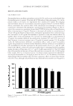
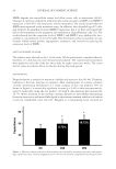
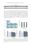
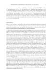
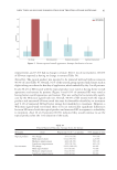
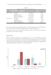
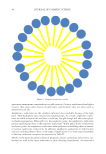
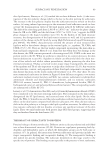
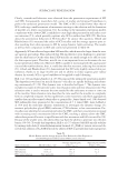
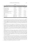
JOURNAL OF COSMETIC SCIENCE 58 RESULTS AND DISCUSSION CELL VIABILITY ASSAY Fu coxanthin has an exce llent antioxidant activity (29,30), and a recent study showed that fucoxanthin protects against Ultraviolet-B (UVB)-induced skin photoaging (23). In the present study, in vitro and in vivo experiments were conducted to evaluate the antiaging and antiwrinkle effects of a fucoxanthin concentrate prepared from P. tricornutum. Before effi cacy assessment, the cytotoxic effect of the concentrate was evaluated at various con- centrations to determine the appropriate concentration for treatment (Figure 2). The re- sults of the cytotoxicity experiment showed that PT-FX50 did not affect cell proliferation when it was used up to 25 μg/ml. However, it decreased cell number at concentrations of 50 and 100 μg/ml. Based on these results, subsequent experiments were conducted using the concentrate at d25 μg/ml. Fucoxanthin is reported to cause few adverse effects on normal cells at a low concentration (31–34). Liu et al. showed that at a concentration of 15 μg/ml, fucoxanthin inhibited the proliferation of glioma U87 and U251 cancer cell lines however, it was not cytotoxic up to 30 μg/ml to normal neurons (31). In another study, the half-maximum inhibitory concentration (IC50) values of pure fucoxanthin against the proliferation of human dermal fi broblasts, Human umbilical vein endothelial cell, and HEK293 cells after treatment for 48 h were found to be 32, 6.7, and 18.7 μM, respectively. The IC50 values of Undaria pinnatifi da extract containing 60.8% fucoxanthin against the three cell lines were 46.5, 6.7, and 33.9 μM, respectively (32). The 50% lethal dose of fucoxanthin in Institute of Cancer Research mice is reported to be 2,000 mg/kg body weight however, no obvious toxicity was observed after repeated oral administra- tion of pure fucoxanthin at 750 mg/kg to mice for 28 d (17,35). In addition, it has been demonstrated that fucoxanthin does not have a genotoxic effect on mouse bone marrow cells (36). Thus, although cell proliferation was inhibited by fucoxanthin at 50 μg/ml in Figure 2. Cytotoxicity of PT-FX50 against HDFN cells. Data are expressed as mean value ± standard deviation (n = 3).
Purchased for the exclusive use of nofirst nolast (unknown)
From: SCC Media Library & Resource Center (library.scconline.org)