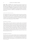
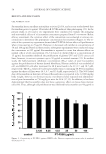
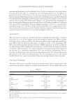
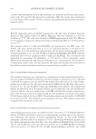
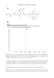
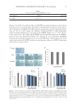
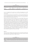
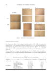
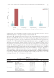
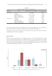
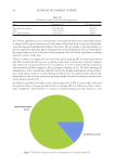
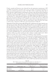
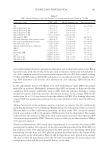
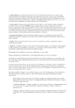
WHITENING AND PROTECTIVE EFFECT OF ALACELL 71 (Figure 2A and B). To study the effect of ALACELL on melanin formation, the melanin formation of 10, 20, and 40 μM on ALACELL-treated B16F10 melanoma cells was quan- tifi ed. Treatment of α-MSH (100 nM) induced a signifi cantly increase of melanin forma- tion (137.2%, p 0.001 Figure 2C). Arbutin (40 μM) and PTU (75 μM) signifi cantly reduced melanin formation in cells induced by α-MSH (Figure 2C p 0.01 and p 0.001, respectively). Five-ALA and ALACELL decreased melanin formation in α-MSH– induced cells, dose dependently (Figure 2C). Among the two compounds, ALACELL decreased more melanin formation at 10, 20, and 40 μM than 5-ALA, respectively. These results indicate that ALACELL exhibits antimelanogenic effi cacy in B16F10 melanoma cells without cytotoxic effect. Figure 2. The cytotoxicity by treatment of ALACELL (A and B), inhibitory effect of ALACELL on melanin formation (C), and inhibitory effect of ALACELL on tyrosinase activity in B16F10 cells (D). B16F10 cell s were treated with concentrations of 10, 20, and 40 μM. After incubation of 48 h, the cells were photographed under a microscope of 2.5 × 10⁴ magnifi cations, and cytotoxicity was measured by the MTT assay. Data are displayed with mean ± standard deviation (n = 3). Signifi cance was indicated at *p 0.05, **p 0.01, and ***p 0.001. Table I The IC50 o f 5-ALA and ALACELL in C. acnes Concentration 5-ALA ALACELL IC50 (mM) 18.01 ± 0.3 6.17 ± .4.0

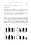
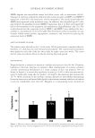
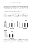
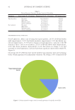
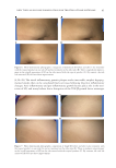
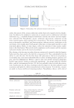
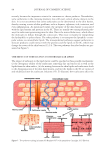
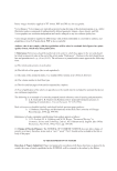
JOURNAL OF COSMETIC SCIENCE 72 ALACELL INHIBITS TYROSINASE ACTIVITY WITHOUT CYTOTOXICITY I N B16F10 CELLS Five-ALA and ALACELL (10, 20, and 40 μM) signifi cantly inhib ited intracellular tyrosi- nase activity as compared with B16F10 melanoma cells treated with α-MSH. In addi- tion, the ALACELL and 5-ALA were shown to reduce tyrosinase activity by 115.6% and 135.4% at 40 μM, respectively (Figure 2D, p 0.001). ALACELL IMPROVES CELL VIABILITY IN UVB-IRRADIATED HaCaT CELLS T o evaluate whether ALACELL protected HaCaT keratinocyte cells against UVB-induced cell death, we detected the viability of HaCaT cells after being exposed to UVB (40, 80, and 120 mJ/cm2) and incubation for 24 h, with and without ALACELL treatment at concentrations of 20, 40, 80, and 100 μM. As shown in Figure 3, the viability of HaCaT cells was decreased signifi cantly to 88.4% after being exposed to UVB (40 mJ/cm2) and for 24 h. 5-ALA did not display recovered viability in UVB-irradiated (40, 80, and 120 mJ/cm2) Figure 3. Protective effec t of ALACELL by 40 mJ/cm2 (A), 80 mJ/cm2 (B), and 120 mJ/cm2 (C) irradiation on UVB-irradiated HaCaT cells. The HaCaT cells were seeded on six-well plates at 7 × 10⁴ cells/well and treated with 40, 80, and 120 mJ/cm2 of UVB for 1 h. The cells were treated with 20, 40, 80, and 100 μM of ALACELL for 12 h, and then followed by UVB treatment. After 24 h, cell viability was measured by the MTT assay. Data are displayed with mean ± standard deviation (n = 3). Signifi cance was indicated at *p 0.05, **p 0.01, and ***p 0.001.
Purchased for the exclusive use of nofirst nolast (unknown)
From: SCC Media Library & Resource Center (library.scconline.org)