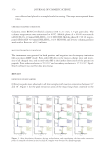
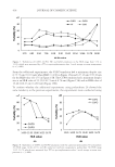
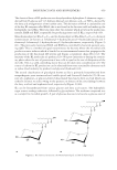
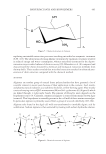
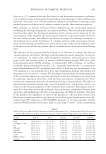
CERAMIDE IN SKIN AND BLOOD 369 Apparatuses . Q Exactive quadrupole-electrostatic fi eld track well high-resolution FT MS machine and Ultimate 3000 HPLC instrument were obtained from Thermo Fisher Scien- tifi c Company (Waltham, MA), Heraeus Fresco 17 Centrifuge and Vacuum freeze dryer were provided by Shanghai Thermo Fisher Biotechnology Company Limited, JK-100B Ultrasonic Cleaner was purchased from Hefei Jin Nick Machinery Manufacturing Com- pany Limited (Hefei,China), Millex-HV needle type fi lter was obtained from Shanghai Hehe Technology Company Limited (Shanghai,China), and EYELA rotary evaporator was from the N-1300 series Tokyo Physical and Chemical Equipment Company Lim- ited (Tokyo, Japan). PRETREATMENT OF SKIN SC SAMPLES SC sample acquisition. A thin layer of cyanoacrylate-containing glue was coated onto one end of a slide. A 2.5 × 2.5-cm area close to the volar side of the forearm was prepared. A force of 1 newton was used to press the slide to the forearm for 1 min followed by carefully stripping. This was repeated at the same site for three times. D issolution. Epidermis on the slides was placed in a 100-ml beaker followed by immersion in 5 ml of chloroform and 25 μl of methanol (chloroform:methanol, 99.5:0.5, v:v). A fter mixing well, the stripped corneocytes in chloroform methanol were placed in JK-100B ultrasonic cleaner and subjected to ultrasonic (100 W) shock for 20 min until fully dissolved. Fi ltration. The above suspension was then transferred to a round-bottom fl ask using a syringe and a 0.22-μm fi lter. Dr ying. The fl ask was connected to a rotary evaporator and evaporated at 40°C until the liquid evaporated. Red issolution. One milliliter of methanol was added to the fl ask, mixed well, and then the liquid was transferred to a 2-ml centrifuge tube. Cent rifuge. The sample was subjected to centrifugation (2,000 r/min) for 10 min at 4°C and the supernatant transferred to a new 2-ml centrifuge tube, followed by testing. PRETR EATMENT OF BLOOD SAMPLES (i) Two hundred microliters of the blood sample was transferred to a 15-ml centrifuge tube. (ii) Then, 1.5 ml of methanol was added into the centrifuge tube and vortexed until fully mixed. (iii) Five milliliters of isopropanol was added into the centrifuge tube and vortexed. (iv) Then, 1.25 ml of ultrapure water was added to the centrifuge tube and vortexed. (v) The mixture was then allowed to stand for 15 min at about 23°C to separate into layers. The supernatant (isopropanol layer) was taken for analysis. (vi) T he supernatant was freeze-dried with a vacuum freeze dryer. Then, 200 μl of a 1:1 mixture of isopropanol and acetonitrile was added and centrifuged. The supernatant

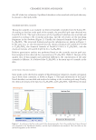
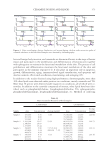
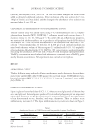
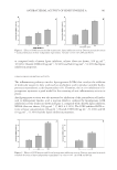
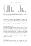
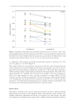
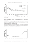
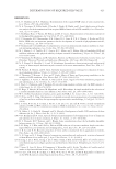
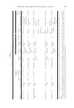
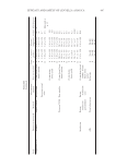
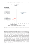
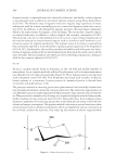
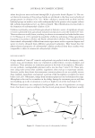
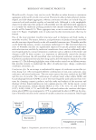
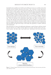
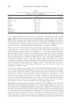
JOURNAL OF COSMETIC SCIENCE 370 was collected and placed in a sample bottle for testing. The steps were repeated three times. CHROMAT OGRAPHIC CONDITIONS Columns were BEH C18 Shield columns (100 × 2.1 mm, 1.7-μm particles). The column temperature was maintained at 40°C. Mobile phase A = 60:40 acetonitrile (ACN)/H2O + 10 mmol NH4HCO2, 0.1% HCOOH Mobile phase B = 90:10 isopro- panol (IPA)/ACN +10 mmol NH4HCO2, 0.1% HCOOH, and reverse column gradient wash with a fl ow rate of 0.3 ml/min. MASS SPECTR OMETRIC CONDITIONS The instrum ent was operated in both positive and negative ion electrospray ionization full-scan mass ddMS2 mode. First-order MS detects the mass-to-charge ratio and inten- sity of all charged ions, and second-order MS is the further dissociation of the parent ion peptide. First-order resolution is 70 × 103 and secondary resolution is 17.5 × 10³. Lipid- Search software was used for data processing. RESULTS DETECTI ON OF SK IN SAMPLES Q Exactive peak s were observed in all skin samples with reaction intensities between 105 and 107. Figure 1 list the peak extraction areas of the major long-chain ceramide in the Figure 1. After dissolution, fi ltration, drying, redissolution, and centrifuging, the fi rst-order extraction peaks of ceramide subclasses in the four skin SC samples were obtained by chromatography.
Purchased for the exclusive use of nofirst nolast (unknown)
From: SCC Media Library & Resource Center (library.scconline.org)