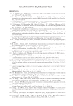
SOOTHING EFFECT OF POGOSTEMON CABLIN EXTRACT 427 This process allowed us to obtain a nonpolar fraction (containing essential oil), and a polar fraction containing molecules such as phytosterols, fl avonoids, and fatty esters. Pure extract was diluted in octyldodecanol, and was used at 1% on ex vivo skin and at 0.001% on cultured keratinocytes. ANTIBODI ES Primary antibodies used were anti-CNR2 (Thermo Scientifi c, Waltham, MA), anti–β- endorphin (LSBio, Seattle, WA), anti–interleukin receptor 1 (IL1R1) (Rockland, Limerick, PA), anti–interleukin 6 signal transducer (IL6ST) (Santa Cruz Biotechnology, Heidelberg, Germany), and anti-TRPV1 (Thermo Scientifi c). Alexa Fluor® coupled secondary anti- bodies were used (Molecular Probes, Eugene, OR). REAGENTS Sel ective an d competitive antagonists of cannabinoid receptor 2: AM630 and selective agonist of cannabinoid receptor 2: AM1241 were purchased from Sigma Chemical Co. (St. Louis, MO). AM630 and AM1241 were applied on skin biopsies at 1 mM, 3 h (topical and culture medium application) before UVB irradiation with 200 mJ/cm2 (Bio-Link Irradiator 254 nm, Fisher Scientifi c, Illkirch, France). Lipopolysaccharide (LPS) (Sigma- Aldrich, St. Louis, MO) was used at 0.5 mg/mL overnight. CELL CULTURE Normal human epithelial keratinocytes were isolated from skin obtained from plastic surgery of healthy females who had given written informed consent. Keratinocytes were cultured in keratinocyte serum–free medium, with provided human recombinant epider- mal growth factor and bovine pituitary extract (Gibco, Auckland, New Zealand), and 0.1 mg/mL Primocin™ (Invivogen, San Diego, CA). β-ENDORPHIN S YNTHESIS IN CULTURED KERATINOCYTES Human keratino cytes were grown in tissue culture dishes (100 mm). After the addition of the patchouli extract, plates were incubated for 24 h with the patchouli extract at 0.001% [diluted in 0.001% dimethyl sulfoxide (DMSO)]. Cells were harvested, and β-endorphin was measured by enzyme-linked immunosorbent assay (Elabscience, Houston, TX). HEMOTOXYLIN AND EOSIN STAINING Slides containi ng 4 μm skin paraffi n sections were deparaffi nized and rehydrated in the following baths: 2 × 2 min in xylene, 2 × 2 min in 100% ethanol, 1 × 2 min in 95% ethanol, 1 × 2 min in 80% ethanol, and 1 × 5 min in H2O. Hematoxylin staining: 1 × 3 min hematoxylin and rinsed in water for 1 × 5 min. Eosin staining and dehydration:

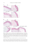
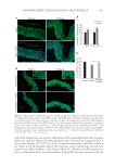
JOURNAL OF COSMETIC SCIENCE 428 1 × 2 min in eosin, 1 × 2 min in 95% ethanol, 2 × 2 min in 100% ethanol, and 2 × 2 min in xylene. Coverslips were mounted on glass slides using Eukitt® (xylene based) as mount- ing media (O. Kindler, Germany) and dried overnight. IMMUNOHISTOLOGICAL FLUORESCENCE Human sk in samples were obtained from pl astic surgery of healthy females who had given written informed consent. After removal of subcutaneous fat, the tissue was used to obtain 6 mm punch biopsies that were incubated with the patchouli extract (or its pla- cebo) at 1% for 48 h. Biopsies were then fi xed in formaldehyde and processed in an auto- mated Shandon Hypercenter XP (Shandon Ltd., Runcor, United Kingdom) for paraffi n embedding. Sections of 4 μm thickness were cut with a microtome (Shandon) and col- lected on polylysine-coated glass slides (Menzel Gläser, Braunschweig, Germany) for im- munohistochemistry. Heat and pepsin enzymatic antigen retrieval was performed before incubation with CB2 receptor and β-endorphin antibodies. Heat antigen retrieval was performed before incubation with IL1R1 primary antibody. Pepsin enzymatic antigen retrieval was performed before incubation with TRPV1 primary antibody. No antigen retrieval was performed before incubation with IL6ST primary antibody. After incubation with the secondary antibody, cell nuclei were stained with 4′,6-diamidino-2-phenylindole (DAPI). Skin sections were viewed under a microscope (Axiovert 200M, Carl Zeiss, Oberkochen, Germany) and photographed with a CCD camera (EXI blue, Qimaging, Surrey, BC). SUBJECT To demonstrate the potential effect of patch ouli extract on sensitive skin, a stinging test was performed as the reference test for sensitive skin. However, other parameters known to be altered on sensitive skin, like skin barrier function, were evaluated on these 26 volunteers on the forearm at the same time as the stinging test (data not shown). One month before the beginning of the clinical test, panels of subjects who are sensitive to 0.01% of capsa- icin diluted in 10% ethanol (which cause sensory irritation) and not to 10% ethanol, applying on nasolabial fold during 10 s, were preselected by a trained expert. Just after the exposure, the subject recorded the sensations of stinging each minute during 10 min on a 0- to 4-interval scales (0 = no sensation, 1 = slight stinging, 2 = moderate stinging, 3 = intense stinging, and 4 = very intense stinging). At the end of the 10 min, the 11 values recorded by the subject were summed up to a fi nal score. If this score was superior to 10, the volunteer was declared like having sensitive skin. Only volunteers having a score superior to 10 were identifi ed as having sensitive skin and were enrolled in the study. At the beginning, 26 volunteers were enrolled in the study and were divided in two groups of 12 volunteers homogenous in age and gender to be sure that these two parameters did not infl uence the results. However, during the test period, six volunteers had to be excluded of the stinging test results, because of big variation in their stinging sensation compared to other trials done at the same period. For fi ve of these volunteers, the variation was explained by a bad cold, and for the last one, no explanation was found. Thus, the stinging result was performed on 20 volunteers, divided in two groups of 10 volunteers homogenous in age and gender.
Purchased for the exclusive use of nofirst nolast (unknown)
From: SCC Media Library & Resource Center (library.scconline.org)