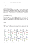
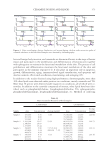
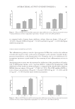
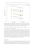
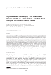
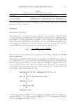
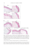
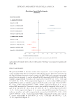
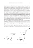
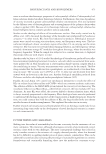
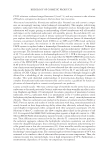
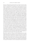
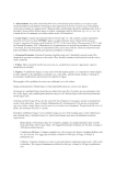
SOOTHING EFFECT OF POGOSTEMON CABLIN EXTRACT 431 cells of the dermis were also positive. Basal layers of the epidermis showed the strongest fl uorescent intensity (Figure 2A). The patchouli extract increased the expression level of the receptor (Figure 2C +22% *p 0.05). A downstream signal of the CB2 receptor is the release of the β-endorphin opioid. The patchouli extract signifi cantly increased the protein level of β-endorphin in skin (Figure 2B and C). Moreover, the β-endorphin re- lease could be observed in the cell interspaces (Figure 2B close-ups) compared with the Figure 2. Expression of cannabinoid receptor 2 and β-endorphin in normal skin. (A) Immunohistochemistry of cannabinoid r eceptor 2 (green) and DNA stained with DAPI (blue), 48 h after topic application of placebo or patchouli extract (×40 objective lens, scale bar = 16 μm). (B) Immunohistochemistry of β-endorphin (green) and DNA stained with DAPI (blue), 48 h after topical application of placebo or patchouli extract (×20 objective lens, scale bar = 31 μm). (C) Quantifi cation of the cannabinoid receptor 2 and β-endorphin immunohistochemistry. (D) Detection of β-endorphin expression in keratinocytes by ELISA, 24 h after patchouli extract addition in the culture media. (n = 3 mean ± Standard Error of the Mean ***p d 0.005 **p d 0.01 *p d 0.05).

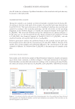
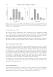
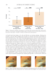
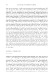
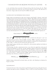
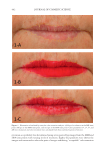
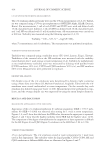
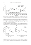
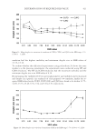
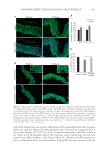
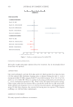
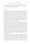
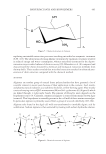
JOURNAL OF COSMETIC SCIENCE 432 placebo condition. Synthesis of β-endorphin by keratinocytes was confi rmed by ELISA (Figure 2D) keratinocytes expressed more β-endorphin when treated with the patchouli extract (+17% *p 0.05). The DMSO (DMSO used at 0.001%) showed a little variation in β-endorphin levels compared with control condition. PATCHOULI EXTRACT MODULATES EXPRESSION OF TRPV1, INTERLEUK IN RECEPTOR 1, AND IL6ST The skin expresses an abundance of Transient Receptor Potential (TRP) channels m odu- lating its development, integrity, and function. TRPV1 receives substantial attention as a candidate target for pain control, and itching sensation in skin. Cannabinoids have been used to treat pain for millennia we tested the hypothesis that the patchouli extract could decrease the expression of TRPV1 and exerted a peripheral soothing effect. UVB and LPS induced TRPV1 immunohistochemistry in human skin (Figure 3A). The treatment with patchouli extract reduced the expression of TRPV1 in skin in response to UVB- or LPS- induced infl ammation (Figure 3A). IL-1 is a highly active and pleiotropic pro-infl ammatory cytokine. The receptor that mediates all known biologic activities of IL-1 is the IL-1 receptor type 1 (IL1R1). We investigated the capacity of the patchouli extract to reduce the expression of IL1R1. Immunohistochemistry results showed that the patchouli extract reduced IL1R1 expression, in skin infl ammation models induced either by UVB (Figure 3A) or LPS (Figure 3B). Interleukin-6 (IL-6) is a pleiotropic cytokine, with diverse roles in driving chronic infl ammation. IL-6 activities are predominantly exerted through a pro- cess known as trans-signaling that uses a protein called IL6ST for initiating the IL6 signal transmission. The effect of the patchouli extract on the local concentrations of IL6ST in skin was investigated. Results showed the capacity of the extract to reduce IL6 signaling through IL6ST diminution in the epidermis irradiated with UVB (Figure 3A and C). CALMING EFFECTS OF PATCHOULI EXTRACT ON CAPSAICIN-INDUCED FACIAL STINGING IN VOLUNTEERS WITH SENSITIVE SKIN. All 20 subjects reported a stinging/burning sensation afte r application of capsaicin. Twenty-eight d of cream applications, with patchouli extract, resulted in signifi cant lower values for burning/stinging sensations in comparison to the values obtained for the placebo group (Figure 4). On the patchouli extract–treated sides, a signifi cant decrease in the TEWL was observed after 28 d of application compared with the placebo sides, demonstrating that the patchouli extract can improve the skin barrier function (data not shown). DISCUSSION In this study, we highlight the key function of cannabinoi d signaling, to control local immune responses in the human skin. Anti-infl ammatory effects of cannabinoid CB2 receptor activation was observed with the agonist AM1241. Recent data with AM630 were available on rodent skin model (28). Our results add comprehension to the human skin physiology, by providing direct evidence supporting the tight control of the cutane- ous endocannabinoid system, in the cellular response to UVB. Several previous studies showed endocannabinoid system have been involved in major roles in nociception or in- fl ammatory reactions by receptor activation (29,30). We hypothesized that the extract
Purchased for the exclusive use of nofirst nolast (unknown)
From: SCC Media Library & Resource Center (library.scconline.org)