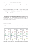
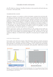
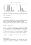
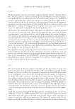
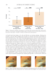
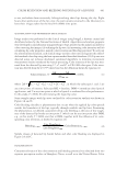
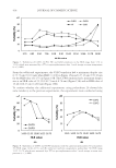
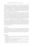
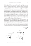
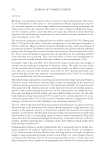
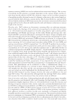
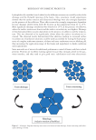
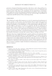
A DEPIGMENTATION TOPICAL TREATMENT PROGRAM ENHANCES A PREVIOUS CHEMICAL PEELING OF THE FACE 395 action, thanks to its glycolic acid content, and a mechanical action, thanks to the presence of squalene and scrubbing beads, specifi cally formulated to help uniform the skin pig- mentation. The glycolic acid contained in the night cream is considered the safest and the most versatile peeling agent among all the alpha hydroxy acids used as chemical peels for the treatment of melasma because it has the smallest molecule and penetrates the epider- mis the best (23). The kojic acid contained in the day and night creams is widely used for melasma treatment as well (24), and its clinically effective antimelanogenic activity on hyperpigmented skin is associated with the induction of Interleukin-6 (IL-6) production in keratinocytes (25), other than with its tyrosinase inhibition and scavenging activity of reactive oxygen species (16). Our dat a revealed no statistically signifi cant change for pigment uniformity and skin texture in the study we performed. Additional investigations with a longer study period and larger sample size will be performed to further verify an effect of the chemical peeling/ depigmentation treatment we tested also on these parameters. This st udy took advantage of the Antera 3D system, which proved to be a valuable, objective, easy, and reproducible method to assess the effects of a dermocosmetic treatment on hy- perpigmentation, excluding any need for clinical scores (26) or self-assessment. The area selected in each patient for the study purposes showed a regular surface, without excessive concavities or convexities, potentially affecting the measurements of the selected skin parameters. Of note, a recent study compared the Antera 3D-based system with other methods (Mexameter® MX-18 and Colorimeter® CL-400, Courage+Khazaka electronic GmbH, Köln, Germany) for the skin color analysis in healthy volunteers. The Antera 3D system sho wed a better sensitivity and specifi city and a higher repeatability versus other methods for all the parameters analyzed (22). The only intrinsic limitation of the Antera 3D system was the fi xed size of the skin areas analyzed (56 × 56 mm). Because th e study product was a home-based treatment, not only its effi cacy but also its safety, tolerability, ease of use, and level of patients’ and physicians’ satisfaction have been considered as parameters of major importance in the assessment of the hyperpigmenta- tion topical treatment program. The treatment was safe and well-tolerated, and the satis- faction rate of investigators and patients with the overall results at study completion was very high. Of interest, a previous multicenter, prospective study performed on 100 women affected by melasma and treated for 45 and 90 d with the same home-based depigmentation pro- gram tested in our study indicated its effectiveness on melasma even in the absence of a prestudy chemical peeling (27). Figure 4. Pie charts sh o wing the distribution of subjects’ opinions on the pleasantness of the study products included in the depigmentation topical treatment program (including the cleanser, day cream, and night cream).

JOURNAL OF COSMETIC SCIENCE 396 In conclusion , our data support the effi cacy, safety, and tolerability of a home-based de- pigmentation treatment based on the combination of different ingredients and activities, leading to a potential synergic effect and enhancing the overall activity of the treatment program. Subjects’ compliance with this home-based daily treatment has been assured by its ease of use and high pleasantness, as rated by the large majority of the study participants. ACKNOWLEDGMEN T S We are gratef ul to Tania Torresani for assistance in clinical activities and to ERA ms Srl for assistance in medical writing. This st udy was sponsored by Relife S.r.l. Dr. Cavallini has been the principal investigator in the clinical study he is a consultant for Relife S.r.l. REFERENCES (1) R. Balkr i shnan, A. J. McMichael, F. T. Camacho, F. Saltzberg, T. S. Housman, S. Grummer, S. R. Feldman, and M. M. Chren, Development and validation of a health-related quality of life instrument for women with melasma, Br. J. Dermatol., 149, 572–577 (2003). (2) T. F. Ce s tari, L. P. Dantas, and J. C. Boza, Acquired hyperpigmentations, An. Bras. Dermatol., 89, 11–25 (2014). (3) E. C. Da v is and V. D. Callender, Postinfl ammatory hyperpigmentation: a review of the epidemiology, clinical features, and treatment options in skin of color, J. Clin. Aesthet. Dermatol., 3, 20–31 (2010). (4) C. G. Ri t ter, D. V. Fiss, J. A. Borges da Costa, R. R. de Carvalho, G. Bauermann, and T. F. Cestari, Extra-facial melasma: clinical, histopathological, and immunohistochemical case-control study, J. Eur. Acad. Dermatol. Venereol., 27, 1088–1094 (2013). (5) O. A. Og b echie-Godec and N. Elbuluk, Melasma: an up-to-date comprehensive review, Dermatol. Ther., 7, 305–318 (2017). (6) S. C. Ta y lor, Epidemiology of skin diseases in ethnic populations, Dermatol. Clin., 21, 601–607 (2003). (7) A. Filon i , M. Mariano, and N. Cameli, Melasma: how hormones can modulate skin pigmentation, J. Cosmet. Dermatol., 18, 458–463 (2019). (8) S. H. Kw o n, Y. J. Hwang, S. K. Lee, and K. C. Park, Heterogeneous pathology of melasma and its clinical implications, Int. J. Mol. Sci., 17, 824 (2016). (9) S. Kumar i , S. Tien Guan Thng, N. Kumar Verma, and H. K. Gautam, Melanogenesis inhibitors, Acta Derm. Venereol., 98, 924–931 (2018). (10) P. E. G r imes, N. Yamada, and J. Bhawan, Light microscopic, immunohistochemical, and ultrastructural alterations in patients with melasma, Am. J. Dermatopathol., 27, 96–101 (2005). (11) H. Y. K a ng, J. S. Hwang, J. Y. Lee, J. H. Ahn, J. Y. Kim, E. S. Lee, and W. H. Kang, The dermal stem cell factor and c-kit are overexpressed in melasma, Br. J. Dermatol., 154, 1094–1099 (2006). (12) E. H. K i m, Y. C. Kim, E. S. Lee, and H. Y. Kang, The vascular characteristics of melasma, J. Dermatol. Sci., 46, 111–116 (2007). (13) E. J. K i m, H. Y. Park, M. Yaar, and B. A. Gilchrest, Modulation of vascular endothelial growth factor receptors in melanocytes, Exp. Dermatol., 14, 625–633 (2005). (14) C. Whee l er-Jones, R. Abu-Ghazaleh, R. Cospedal, R. A. Houliston, J. Martin, and I. Zachary, Vascular endothelial growth factor stimulates prostacyclin production and activation of cytosolic phospholipase A2 in endothelial cells via p42/p44 mitogen-activated protein kinase, FEBS Lett., 420, 28–32 (1997). (15) M. I. R e ndon, D. S. Berson, J. L. Cohen, W. E. Roberts, I. Starker, and B. Wang, Evidence and consid- erations in the application of chemical peels in skin disorders and aesthetic resurfacing, J. Clin. Aesthet. Dermatol., 3, 32–43 (2010). (16) N. Bagh e rani, S. Gianfaldoni, and B. Smoller, An overview on melasma, J. Pigment Disord., 2, 218 (2015). (17) D. E. C a stillo and J. E. Keri, Chemical peels in the treatment of acne: patient selection and perspectives, Clin. Cosmet. Investig. Dermatol., 11, 365–372 (2018). (18) T. Sole y mani, J. Lanoue, and Z. Rahman, A practical approach to chemical peels: a review of fundamen- tals and step-by-step algorithmic protocol for treatment, J. Clin. Aesthet. Dermatol., 11, 21–28 (2018).
Purchased for the exclusive use of nofirst nolast (unknown)
From: SCC Media Library & Resource Center (library.scconline.org)