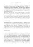
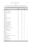
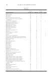
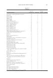
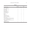
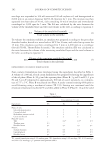
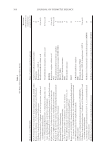
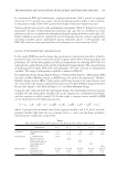
compounds, lipid nanoparticles and NCs are considered colloidal carriers (22). These car- riers allow shielding of chemical compounds against photodegradation phenomena, ensure bioavailability optimization, and allow controlled release, while at the same time, they can be produced in great numbers. These exact colloidal carriers have been proven to amplify the accumulation of UV fi lters on the upper skin layers, as they have been designed to strengthen their photo-protective properties (23). The size of lipid nanocarriers makes skincare products easy to formulate and apply. Melt-emulsifi ed lipids are the base of solid lipid nanoparticle (SLN) formulation, are stable under room-temperature condi- tions, and are made of well-tolerated and biodegradable raw materials (24,25). The h armful effects associated with the exposure to UV radiation are well documented. As a result, the development of a new generation of UV fi lters that can provide effective protection against the entire UV spectrum radiation has become major concern. These protective systems should be carefully designed by selecting substances of highly reliable stability, to ensure optimal safety and effi ciency during the entire time of sun exposure. The a im of this review was to present the methods (in vitro/in vivo) used for the estimation of skin penetration of sunscreens regularly used (some sunscreens listed in both FDA monograph and EU Annex are no longer used because they have an unpleasant feel, irritate skin, or are no longer produced), the studies conducted on their toxicity, the evaluation of margin of safety (MoS), and the current situation and perspectives by using new carriers. IN VI TRO AND IN VIVO METHODS FOR CUTANEOUS PENETRATION AND TRANSDERMAL PERMEATION OF ORGANIC UV FILTERS The s tratum corneum (SC) is the outermost layer of the epidermis. It consists of 10–25 layers of dead, elongated, fully keratinized corneocytes, which are embedded in a matrix of lipid bilayers. Ceramides form the major two-tailed component of the SC lipid matrix. Free fatty acids and cholesterol form the other two dominant components of SC lipids. On most body sites, the SC is 12–16 cell layers thick, but it can vary from as little as nine cell layers on the forehead or eyelids to as much as 25 on the dorsum of the hand and up to 50 or more on the palms or the soles of the feet. The crossing of the SC is the rate-limiting step in the sequence of percutaneous absorption. The desirable site of action of UV fi lters is restricted to the skin surface or within the uppermost layers of the SC. Ideally, a sunscreen should impregnate the SC and create a fi lter against UV radiation, but not penetrate into the underlying viable tissue. Table II UV Filter Substances Categorized According to the FDA GRASEa for use in sunscreens Not GRASEb for use in sunscreens Insuffi cient data for use in sunscreensc ZnO and TiO2 Aminobenzoic acid (PABA) and trolamine salicylate Cinoxate, dioxybenzone, ensulizole, homosalate, meradimate, octinoxate, octisalate, OCR, padimate O, sulisobenzone, oxybenzone, and avobenzone a GRASE = generally recognized as safe and effective. b These ingredients are not currently marketed. c For those ingredients in the “insuffi cient data” category, the FDA proposes that it needs additional data to determine that sunscreens with these ingredients would be GRASE. JOURNAL OF COSMETIC SCIENCE 302

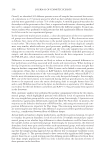
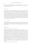
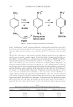
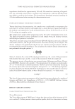
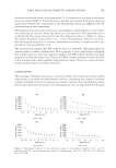
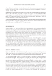
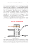
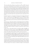
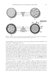
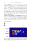
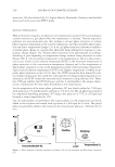
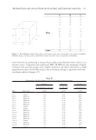
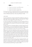
Both i n vivo and in vitro methodologies are available for the evaluation of the skin absorp- tion and percutaneous penetration properties of sunscreen and skincare products. How- ever, in vitro tests are mostly preferred over the in vivo because of ethical reasons and also feasibility. In cases where the crossing of the SC is considered as the foremost rate-limiting step in the process of skin absorption and percutaneous penetration, data come often from in vitro methods. Prediction of in vivo skin absorption and percutaneous permeation, for most of the compounds, is made possible, thanks to the in vitro data deriving from studies using skin membranes. Whereas human skin is only available from surgical sources, excised skin can be easily obtained from animals. This is exactly what makes the production of proper membranes possible, thus opening the way for conducting repro- ducible experiments. With passive diffusion, instead of active, being the cause of penetra- tion, molecular transport is considered the primary route for skin permeation, whereas viability of the skin is not a requirement for penetration testing. In the rare case of dermal biotransformation, it is vital that separated tests are conducted, including fresh excised skin, which could foster a prolong viability under certain circumstances. Test m ethods that are used for the estimation of the in vitro rates, use diffusion cells, also known as Franz cells (Figure 1). These cells consist of an upper and a lower chamber. These chambers are divided by a sample of the human or pig skin in the shape of a disk. Figure 1. Schematic representation of the Franz cell diffusion system (modifi ed from Kim et al.). The donor chamber (1) above the skin contains the applied topical agent. The chamber below the skin is the receptor chamber (2) from which samples are taken through the sampling port. The receptor chamber is surrounded by a water jacket (3) maintained at 32°C. A magnetic stirrer and stirring helix are magnetically rotated at the bottom of the receptor chamber. The topical drug, which is applied to the SC side of the skin, permeates into the dermis side and then crosses the skin (26). DISTRIBUTION OF UV FILTERS ON THE SKIN 303
Purchased for the exclusive use of nofirst nolast (unknown)
From: SCC Media Library & Resource Center (library.scconline.org)