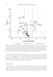
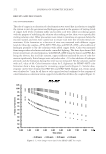
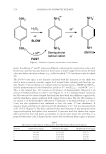
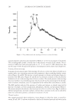
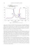
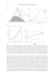
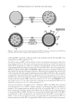
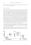
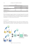
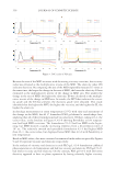
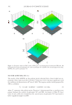
Both i n vivo and in vitro methodologies are available for the evaluation of the skin absorp- tion and percutaneous penetration properties of sunscreen and skincare products. How- ever, in vitro tests are mostly preferred over the in vivo because of ethical reasons and also feasibility. In cases where the crossing of the SC is considered as the foremost rate-limiting step in the process of skin absorption and percutaneous penetration, data come often from in vitro methods. Prediction of in vivo skin absorption and percutaneous permeation, for most of the compounds, is made possible, thanks to the in vitro data deriving from studies using skin membranes. Whereas human skin is only available from surgical sources, excised skin can be easily obtained from animals. This is exactly what makes the production of proper membranes possible, thus opening the way for conducting repro- ducible experiments. With passive diffusion, instead of active, being the cause of penetra- tion, molecular transport is considered the primary route for skin permeation, whereas viability of the skin is not a requirement for penetration testing. In the rare case of dermal biotransformation, it is vital that separated tests are conducted, including fresh excised skin, which could foster a prolong viability under certain circumstances. Test m ethods that are used for the estimation of the in vitro rates, use diffusion cells, also known as Franz cells (Figure 1). These cells consist of an upper and a lower chamber. These chambers are divided by a sample of the human or pig skin in the shape of a disk. Figure 1. Schematic representation of the Franz cell diffusion system (modifi ed from Kim et al.). The donor chamber (1) above the skin contains the applied topical agent. The chamber below the skin is the receptor chamber (2) from which samples are taken through the sampling port. The receptor chamber is surrounded by a water jacket (3) maintained at 32°C. A magnetic stirrer and stirring helix are magnetically rotated at the bottom of the receptor chamber. The topical drug, which is applied to the SC side of the skin, permeates into the dermis side and then crosses the skin (26). DISTRIBUTION OF UV FILTERS ON THE SKIN 303

A reactor fl uid is present in the lower chamber, helping the simulation of the circulation in the dermis. The substance to be tested is placed on the skin, and its diffusion through the skin is evaluated by analyzing the receptor fl uid, which is located in the lower cham- ber. After the diffusion period is terminated, several skin layers are examined for any re- sidual materials. The UV fi lters are likely to exhibit different penetration rates, in contrast to pure substances, because they are examined in standardized preparations, at several concentrations. After application (the duration varies at every experiment), the samples are rinsed using a surfactant solution, to analyze the concentration of the product in every layer (SC, epidermis, dermis, and receptor fl uid). The amount of the applied test sub- stance, which is found in the SC, is estimated by the so-called SC absorption. By dermal absorption, on the other hand, we mean the amount of the applied product found in the epidermis and dermis. Last, by percutaneous absorption, we mean the amount of the ap- plied product that is found in the receptor fl uid. An in vivo method, for measuring skin penetration, is the tape-stripping method. For 30 min, samples are placed on the skin surface, and then, the substance in excess is removed from the surface by swiping with a dry cloth. Seven tape strippings are used to remove the SC of the area under examination. The fi rst tape strip is thrown away, whereas the next six are gathered, put in a beaker, containing a suitable solvent and stirred for 30 min. The levels of the sunscreen product in the solvent are then measured (27). It is well known that there are considerable differences between animal and humans in their skin delivery systems, attributed to factors including SC thickness, hydration, and lipid composition. Using fresh frozen human skin instead of animal skin can serve as an excellent alternative to methods using animal skin while also making the result much closer to human living skin. Although the tissue has been frozen and stored at -80°C, transport and barrier mechanisms apparently remain functional. Similar distribution patterns have also been demonstrated in the porcine skin. Although the experiments are performed with the skin kept at -80°C, freeze–thaw cycles or careless storage at higher temperatures might affect the results and the permeability of the skin. Nevertheless, ethically, it is an excellent way to avoid using experimental animals in permeation studies (28). TOXICITY ST UDIES Scientists are well aware of toxicity issues related to fi lters. This concern has been confi rmed by a number of studies (in vitro/in vivo), according to which commonly used sunscreens were found to have an endocrine active chemical action. In vitro studies investigated estrogenic activity of UV fi lters, varying in their design and endpoints, which might explain the diverging results. Most in vitro studies reported that BP-3, 4-methylbenzylidene camphor (4-MBC), OMC, HMC, and OD-PABA exhibit estrogenic activity. However, not all of the UV fi lters exhibiting in vitro estrogenic activity were estrogenic in acute in vivo models. In vitro, 8/9 c hemicals (BP-1, BP-2, BP-3, 3-BC, 4-MBC, HMS, OD-PABA, and OMC) showed estrogenic (on MCF-7 cells) and 2/9 (BP-3 and HMS) showed antiandrogenic activity (on MDA-kb2 cells). Six/nine fi lters (BP-1, BP-2, BP-3, 3-BC, 4-MBC, and OMC) increased uterine weight in immature rats. 3-BC and 4-MBC displaced 16α125I- estradiol from human estrogen receptor (ER)β. Developmental toxicity of 4-MBC (0.7–47 mg/kg body weight/day) and 3-BC (0.24–7 mg/kg), administered in chow, was investigated in the Long–Evans rats. Weight gain of pregnant rats was reduced only by JOURNAL OF COSMETIC SCIENCE 304
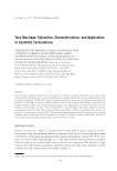
Purchased for the exclusive use of nofirst nolast (unknown)
From: SCC Media Library & Resource Center (library.scconline.org)