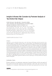
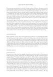
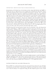
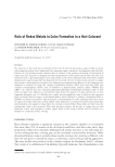
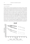
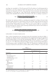
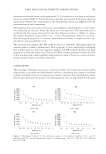
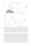
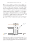
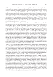
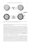
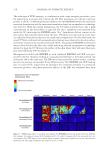
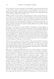
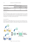
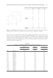
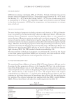
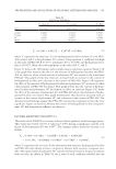
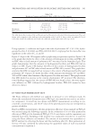
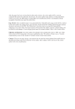
of the high BP-3 partition coeffi cient between the epidermis and the SC when BP-3 was formulated into NLC suspension (76). The effectiv eness of EHT and the ability to limit any possible toxicological effects has been made possible by the restricted percutaneous penetration of lipid microparticles (LMs). EHT encapsulation in LMs on its diffusion through the SC with glyceryl behenate and phosphatidylcholine has been examined. Creams with EHT in free or encapsulated in LM form combined with OMC and BMDBM, which are the most common UV fi lters and were applied on the skin of human volunteers. What was also examined was the fraction of the cream dose that was applied and had permeated in different SC layers. The cream that contained the nonencapsulated sunscreen agent presented a percentage of 21.9 ± 4.9% of the EHT dose, which was diffused in the SC. This percentage did not differ a lot from the smaller molecular weight OMC, which was found at 22.2 ± 7.6%, and BMDBM, which was found at 20.5 ± 3.7%. The cream that contained microencapsulated EHT gave an important reduction in 45.7% in skin permeation (77). Nanostructured Pol ymer and Lipid Carriers. Polymeric (PLC) a nd SLNs were prepared and characterized to act as BP-3 carriers, aiming at optimizing the safety of sunscreen products. The nanoprecipitation method was used to encapsulate BP-3 (1.6% w/w) in poly nanoparticles (epsilon-caprolactone) (PCL) and hot high pressure homogenization, to encapsulate BP-3 in SLNs. In both cases, the particles remained stable for 40 d. The BP-3 encapsulated in PCL nanoparticles was released faster than BP-3 encapsulated in SLNs. A raise in the sun protection factor concurred with the encapsulation of BP-3 in both nanostructures. Also, BP-3, encapsulated in SLNs, did not seem to cause any cytotoxic Figure 2. Schematic representation of lipid nanoparticles [SLNs and nanostructured lipid carrier (NLC)] and lipid NCs NPLC and NCs in aqueous media [modifi ed from Kaul et al. (75)]. DISTRIBUTION OF UV FILTERS ON THE SKIN 315

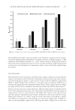
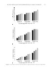
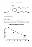
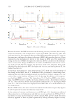
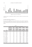
or phototoxic effects in human keratinocytes (HaCaT cells) and BABL/c 3T3 fi broblasts, whereas PCL nanoparticles with BP-3 revealed phototoxic potential in HaCaT cells. The studies of skin permea tion were made over 24 h in a Franz diffusion cell with the human skin, donated from plastic surgery. After this, the BP-3 amounts were measured in epidermis/dermis by the tapping-stripping method, as described in the receptor fl uid by HPLC. During the skin permeation study, it was observed that BP-3 encapsulation in the PCL nanostructure decreased its penetration into the skin. PCL nanoparticles decreased BP-3 skin permeation by 70% in the epidermis and dermis and 80% in the receptor fl uid. However, the skin permeation of SLN-BP-3 was not signifi cantly different from that of free BP-3 (78). Vettor et al., evaluated O MC distribution in skin compartments from OMC-loaded poly-D,L-lactic acid (PLA) nanoparticles formulated in an emulsion gel (OMC-NP emulgel) comparatively to a nonencapsulated OMC emulsion gel (OMC emulgel). Both formulations contained 5% OMC. The classical Franz cell method was fi rst applied, and OMC amounts in each skin strata [SC, epidermis (E), and dermis (D), in the receptor fl uid (RF) and at the skin surface (unpenetrated dose)] were determined after 1, 2, and 3 h exposure time. The results showed the in vitro distribution of OMC in skin compartments for both formulations using either “ACET” (extraction of OMC with acetone) or “IPM” (extrac- tion of OMC with isopropyl myristate) methods. Comparison between OMC emulgel and OMC-NP emulgel gave useful information on the cutaneous uptake of OMC in the skin depending on both formulations. When applied encapsulated in NP, the major part of OMC was retained at the skin surface over time. After 2 and 3 h, 85 and 80% of the applied OMC were, respectively, recovered on the skin surface with NP. These amounts were much higher than those obtained when OMC emulgel was applied (62 and 56%, respectively, after 2 and 3 h). This result demonstrated the nanoparticle ac- cumulation at the skin surface. High amounts of OMC were detected in the SC (be- tween 12 and 26% for OMC emulgel and 5–8% for OMC-NP emulgel with ACET method. More than 80% of OMC conce ntrated on the top of the skin and in the SC after 3 h exposure time. The main difference between the OMC emulgel and the OMC-NP emulgel was OMC distribution between these two compartments. In the case of NP, the percentage of accumu- lated OMC was 10-fold higher on the top of the skin than that in the SC. This value dropped to twofold with OMC emulgel. Consequently, OMC amounts in viable skin layers (Qabs = E + D + RF) were superior for OMC emulgel than for OMC-NP emulgel (~3.5 vs. ~2% after 3 h) because the SC may play a role of reservoir. This result confi rmed the higher affi nity of OMC (lipophilic substance, logp = 5.68) for the lipophilic skin layers, and second that the transport of NP was clearly impeded by the SC (39). Luppi et al. synthesized l ipophilic polymers composed of polyvinyl alcohol (PVA) and various fatty acids (FAs) and investigated in vitro the infl uence of the different nanopar- ticles prepared on percutaneous absorption of BP-3. PVA was selected as a starting material for the preparation of such polymers due to its biocompatibility and the pos- sibility for substitution through chemical linkage to its oxy-residues able to modify its physicochemical properties. PVA was substituted, at two different substitution degrees (40 and 80%), with saturated FAs (myristic, palmitic, stearic, and behenic acid) to give to the polymer suffi cient lipophilicity to allow preparation of nanomatrices for sun- screen delivery. JOURNAL OF COSMETIC SCIENCE 316
Purchased for the exclusive use of nofirst nolast (unknown)
From: SCC Media Library & Resource Center (library.scconline.org)