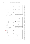
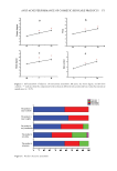
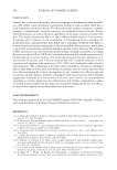
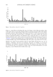
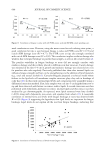
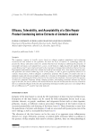
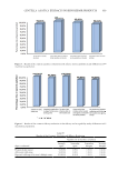
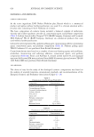
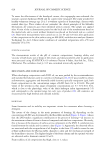
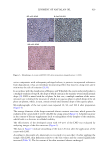
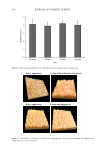
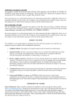
587 SUPPRESSION OF ITCHING BY THREE HERBAL ETHANOLIC EXTRACTS Figure 2. Effect of the ethanolic extracts of three selected herbs on cytotoxicity in HaCaT cells. HaCaT cells were treated with different concentrations (0–80 μg/mL) of A houstonianum, B falcatum, and S chinensis for 24 h (A) or a combination (40 μg/mL A houstonianum, 20 μg/mL B falcatum, and 40 μg/mL S chinensis) for varying lengths of time (0–48 h) (B). Cell viability was measured using a commercial water-soluble formazan-based assay kit (Cell Counting Kit-8). Figure 1. Analytical HPLC profiles of authentic standard and ethanolic extracts. Chromatograms of ethanolic extract of A houstonium (A), ethanolic extract of B falcatum (B), and ethanolic extract of S chinensis (C). Profiles of authentic standard compounds (STD 0.1 mg/mL) were plotted in the upper panel.

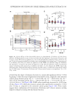
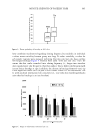
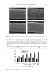
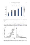
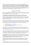
588 JOURNAL OF COSMETIC SCIENCE pro-FLG, which is proteolytically cleaved and converted to FLG (35). FLG is a filament- associated protein that plays an essential role in maintaining epidermal homeostasis (e.g., epidermal hydration and skin barrier function) (36,37). FLG deficiency is highly associated with TEWL, dryness, and bacterial colonization, which confers susceptibility to itch (38– 41). Besides inherited loss-of-function mutations, several inflammatory cytokines, including IL4 and IL13, can induce the suppression of FLG expression at the transcriptional level in AD (20,42,43). Therefore, it has been suggested that restoration of reduced FLG expression by inflammatory cytokines may help relieve itchy skin. Here, we confirmed that treatment with IL4 + IL13 downregulated FLG mRNA expression in a time-dependent manner, as revealed by RT-PCR (Figure 3A) and qPCR (Figure 3B). IL4 + IL13 decreased FLG mRNA levels by 0.98 ± 0.015-, 0.87 ± 0.045-, 0.71 ± 0.036-, and 0.48 ± 0.076-fold increases at 6, 12, 24, and 36 h, respectively, of the control. Consistent with mRNA levels, a time-dependent decrease in pro-FLG protein levels by IL4 + IL13 stimulation was observed using immunoblot analysis (Figure 3C). To Figure 3. Effect of the three herbal extracts on the restoration of IL4 + IL13-induced suppression of FLG expression in HaCaT cells. (A and B) HaCaT cells were treated with IL4 (20 ng/mL) plus IL13 (50 ng/mL) for various time periods (0–24 h). Total RNA was isolated, and FLG mRNA levels were examined using RT-PCR (A) and qPCR (B). GAPDH was used as the loading control. (C) HaCaT cells were treated as in (A), and whole- cell lysates were immunoblotted using anti-FLG antibodies. GAPDH was used as the loading control. (D and E) HaCaT cells were treated with IL4 (20 ng/mL) plus IL13 (50 ng/mL) for 36 h in the absence or presence of A houstonianum (40 μg/mL), B falcatum (20 μg/mL), S chinensis (40 μg/mL), or a combination of the three. Total RNA was isolated, and FLG mRNA levels were examined using RT-PCR (D) and qPCR (E). GAPDH was used as the loading control. (F) HaCaT cells were treated as in (D), and whole-cell lysates were immunoblotted using anti-FLG antibodies. GAPDH was used as the loading control. Relative band intensities were measured using ImageJ. ns: not significant *p 0.05, **p 0.01, ***p 0.001 compared to control (n = 3).
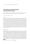
Purchased for the exclusive use of nofirst nolast (unknown)
From: SCC Media Library & Resource Center (library.scconline.org)