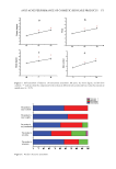
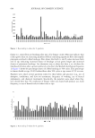
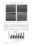
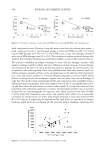
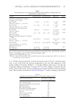
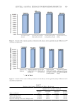
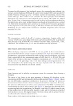
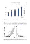
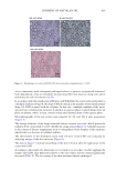
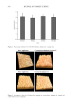
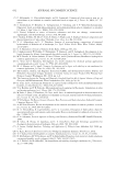
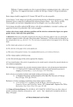
607 FATIGUE STRENGTH OF PANELIST HAIR color decreased (i.e., as color frequency increased cuticle damage also increased). Cuticle removal increases as consumers color more frequently because oxidative damage increases and more disulfide bonds and other protein bonds are broken. Hair subjected to extensive oxidative damage has also been previously shown to have higher propensity for physical damage (9). These two well-established damage measures were then correlated with fatigue breakage data (Figure 8) for tips. SEM grading was not performed at roots and FTIR showed a very Figure 6. Example images for SEM grading scale. (A) Low damage, (B) medium damage, (C) high damage, (D) stripped. Figure 7. SEM cuticle damage score versus time since last color.

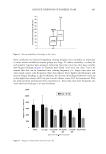
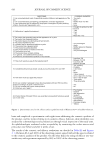
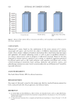
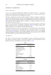
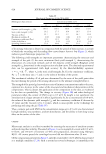
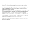
608 JOURNAL OF COSMETIC SCIENCE weak correlation at roots. However, using the mean scores for each coloring time point,, a good correlation for tips is seen between fatigue α-value and FTIR score (R2=0.73) and cuticle SEM damage score (R2=0.77). The FTIR score at tips also strongly correlated with cuticle SEM damage score (R2=0.91). This correlation implies oxidative damage also weakens hair to fatigue breakage in panelist hair samples, as seen in lab created tresses (2). The panelist variability in fatigue breakage at roots did not strongly correlate with oxidative damage and this is likely related to differences in hair structure. Cuticle damage was minimal in the first 6–8 cm of growth and oxidative damage was relatively low even for panelists who color regularly. Previously published data show that internal lipids can influence fatigue strength and hair can be strengthening via the addition of lipid materials (e.g., cetyl and stearyl alcohol) (3). Camacho-Bragado proposed a technical model where failures in the lipid-rich cell membrane complex structure play a key role in breakage of curly hair (10). In this study, internal lipid levels and composition were quantified using a differential extraction method. First, hair samples were cut into 5 cm lengths from root to tip and then extracted with hexane to remove surface lipids. A second extraction was then performed with chloroform–methanol to remove internal lipids and this extract was then analyzed by gas chromatography. As expected, most lipids extracted were fatty alcohols (∼80%) along with cholesterols, wax esters, and squalene from sebum (11). A reasonable correlation in internal fatty acid levels with fatigue breakage at roots was observed with R2=0.36 (Figure 9) supporting the hypothesis that lipid levels are important for fatigue breakage. Lipid levels do not explain all the root hair fatigue breakage, indicating that Figure 8. Correlation of fatigue α-value with (A) FTIR cysteic acid and (B) SEM cuticle grading score. Figure 9. Correlation of internal fatty acid levels versus fatigue α-value.
Purchased for the exclusive use of nofirst nolast (unknown)
From: SCC Media Library & Resource Center (library.scconline.org)