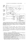
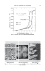
16 JOURNAL OF THE SOCIETY OF COSMETIC CHEMISTS fibers and the disintegration of structures which apparently do not directly contribute to the formation of keratin, such as the nucleus and some parts of cytoplasm. Keratinization of the epidermal cells results in the formation of a so- called soft keratin, which flakes off and is characterized by a relatively low cystine content. Very little is known about the chemical changes that occur during the synthesis of epidermal keratin. The scant analytical data on the amino acid composition of human epidermis and horny layer are listed in Table 1. TABLE I--AMINO ACID COMPOSITION OF HUMAN EPIDERMIS AND HORNY LAYE• Epidermis Horny layer Arginine Lysine Histidine Cystine Tyrosine•yptophane 6.7 5.9 2.6 1.4 4.0 1.0 5.9-10.0 3.1-7.1 0.6-1.9 2.3-3.8 3.•5.7 0.5-1.8 These data, originating from different authors (1, 10), are open to some criticism. "Epidermis" in the table stands for whole epidermis which comprises both cellular and keratinous components, while the horny layer analyzed represents material from a variety of normal and pathological sources. Moreover, methodological and biological variations may account for the variability of the results. With these reservations in mind, it would appear that during keratinization there is little change in the over-all amino acid composition, except for a possible reduction in the histidine con- tent and an increase in the concentration of cystine. In our own analyses of the horny and cellular layers from the same samples of human epidermis, the cystine values remained essentially the same, or showed a slight increase only, throughout the entire thickness of the epidermis (6, 11). This finding is of some theoretical interest. In the past, on the basis of inadequate histochemical evidence, it has been claimed that the oxidation of sulfhydryl groups to disulfide linkages played a major role in epidermal keratinization (12). This theory could not be confirmed either by recent histochemical studies (13) or by our direct chemi- cal analyses (6, 11, 14). The constancy of the cystine values throughout the entire thickness of the epidermis suggests that the consolidaton of the keratin fibers through disulfide bridge formation starts already in the deep cellular layers of the epidermis, and that there is no extensive disulfide bridge formation between the cellular and horny layers, as has been pre- viously thought. Of interest is also the recent finding that sulfhydryl groups (cysteine) are retained in all keratins studied, especially in epidermal keratins (6, 11). With an increasing rate of keratin formation, the concentration of these groups rises. The persistence of the sulfhydryl groups in the horny layer probably renders keratin more susceptible to chemical and physical in- fluences.

NEWER ASPECTS OF EPIDERMAL DIFFERENTIATION 17 The disintegration of cellular proteins goes hand in hand with keratin formation. The morphologic aspects of this disintegration have been mentioned previously. One of them, the frequent, though by no means constant, appearance of a zone of intensified sulfhydryl staining under- neath the horny layer, the so-called keratogenous zone, has been at- tributed to the unmasking of groups previously hidden inside the protein molecule. The end products of the decomposed proteins ascend with the keratin fibers to the surface of the skin from where they can be extracted and identified. Some of these end products, notably the free amino acids, perform an important physiologic function by giving part of the buffering power to the skin surface and thereby assisting in the neutralization of acids and alkalies applied on the outside of the skin (1). The horny layer, end product of epidermal differentiation, is not of uni- form composition, consisting mainly of keratin, but containing the end products of the broken cellular proteins as well. A number of protein fractions, including globular, mucous, and keratinous ones, were identified in calluses (4). In addition, the epidermis is also an organ for the excretion of endogenous and exogenous substances. In the former group belong the so-called melanoids, derivatives of melanin pigments. The excreted pre- cursors of melanin pigments ("premelanin") may reduce metallic salts to free metals on the skin surface (15). The number of exogenous sub- stances, discarded through the epidermis, is practically limitless. Some of these, such as arsenicals, may become incorporated into the keratin mole- cule and thereby may modify the process of keratinization others, like atabrine, may stain the epidermis and provide a screen against the effects of ultraviolet irradiation. The horny layer, though metabolically inactive, can be subjected to a number of chemical and physical reactions. The majority of cosmetic preparations act on the surface only, by modifying it through physical or chemical mechanisms. Absorption of topical agents, though often stressed by the cosmetic industry, is of minor importance for achieving the desired cosmetic effects. The foregoing discussion leaves little doubt that there are great gaps in our understanding of epidermal differentiation. The recent development of new methods for the study of human epidermal proteins offers promise to solve some of these questions. There is little excuse for the continued exclusive study of specimens obtained from animal sources and even less for implying that the results obtained are applicable to human skin. The in- vestigation of material like the cow's nose (7) or the horse burr (12) may be convenient because of the thickness of the epidermis and the relative avail- ability of these tissues nevertheless, recent progress in techniques, facilitating the study of human epidermal proteins, makes research in this area not only practicable, but imperative. These new methods include:
Purchased for the exclusive use of nofirst nolast (unknown)
From: SCC Media Library & Resource Center (library.scconline.org)