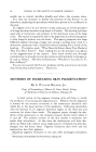
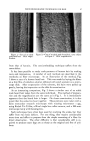
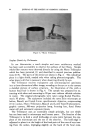
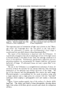
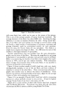
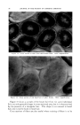
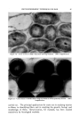
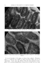
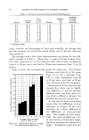
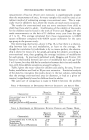
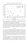
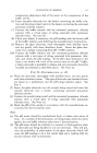
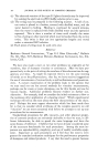
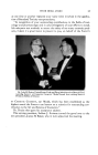
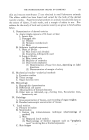
JOURNAL OF THE SOCIETY OF COSMETIC CHEMISTS Figure 3.--Munn Trilineator. Surface Details by Trilineator In our laboratories a much simpler and more satisfactory method has been used successfully to observe the surfaces of hair fibers. Simple whole fiber mounts were observed directly by using the Munn Trilineator which has been patented (5) and described in several previous publica- tions (6-8). The parts of the device are shown in Fig. 3. The cylindrical piece is a light shield, needed only when taking photomicrographs. The other piece is all that is necessary when observing directly by eye. The Trilineator resolves transparent and semitransparent specimens into a three-dimensional effect image, revealing to the eye or to the camera a detailed picture of surface structure. An illustration of this with a human head hair is shown in Fig. 4. The sample was prepared by ex- tracting with ether and mounting in 20 per cent calcium chloride solution in water. The original photographs were taken using Kodak Panchro- matic M plates. The equipment included the same Zeiss microscope as before, Bausch and Lomb 8-mm. apochromatic objective, compensating (15X) ocular, Munn Trilineator, Bausch and Lomb Type H photomicro- graphic camera, 500-watt projection lamp, focusing lens, fused Pyrex water cell and automatic exposure timers. The Trilineator uses an essentially simple principle, but one which has not been used in microscopy until recent years. The function of the Trilineator is to hold a small knife-edge at some point between the eye- piece of the microscope and the eye of the observer. The knife-edge is adjusted to place it at the height of the focal point of the cone of rays issu- ing from the ocular, impinging slightly on the back of the focal cone.

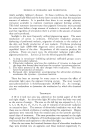
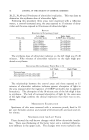
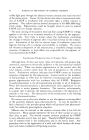
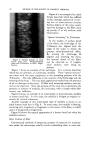
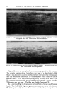
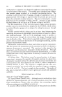
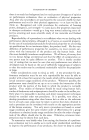
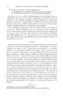
PHOTOMICROGRAPHIC TECHNIQUES FOR HAIR 89 Figure 4.--Surface structure of adult head hair. Photomicrograph taken with Trilineator at 500X magnification. .. 5:. .•:. Figure 5.--Surface structure of child's head hair. Photomicrograph taken with Trilineator at $00X magnification. This important point of intersection of light rays is known as the "Rams- den circle," the "Lagrange disc," the "eye point" or the "exit pupil." When the adjustment is correct, the two-dimensional visual image is transformed into one which shows a three-dimensional effect. The nature of the illumination is important with the Trilineator. It is preferably arranged so that the iris diaphragm in front of the light source is focused sharply in the field of view when the substage condenser is in focus on the specimen. Furthermore, apochromatic objectives and com- pensating eyepieces are recommended for sharper definition and greater perfection of image. If achromatic lenses are used, a green filter is rec- ommended. The use of the Trilineator is a straightforward technique of direct ob- servation without the necessity for any special, elaborate, time-consuming preparation of the specimen and without distortion of the image. The eye of the observer and the photographic film receive the same image. Photomicrography is accomplished by the usual procedure, using only a single exposure and a single print, by contrast with the "bas-relief" technique which employs a photographic trick and renders a distorted image. Figure 5 shows another typical view of the surface of a female child's head hair. The clarity and detail of these photographs, showing almost half of the surface of the fiber, permit accurate scale counts for comparison of fibers.
Purchased for the exclusive use of nofirst nolast (unknown)
From: SCC Media Library & Resource Center (library.scconline.org)