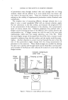
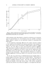
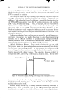
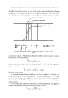
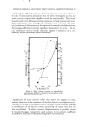
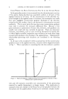
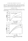
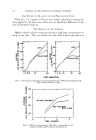
66 JOURNAL OF THE SOCIETY OF COSMETIC CHEMISTS When the skin is thick and the applied material penetrates slowly, the dermis may serve as a reservoir and there may, therefore, be a very long delay before any of the material applied to the cutaneous surface can reach the fluid of the receptor system. It is possible to separate the epidermis and the dermis of nonhairy skins. Flesch (20) studied penetration through the epidermis, as distielct from whole skin, by separating this layer from the dermis after the penetrating substance had been in contact with the epidermal surface for various periods of time. He then detected the presence of the a•l•lied substance at the derreal-epidermal junction. In a refinement of this method, Blank and Gould (14) removed the stratum comeurn from the skin by several strippings with pressure-sensitive tape, then separated the remaining portion of the epidermis from the dermis and quantitatively determined the amount of material which had penetrated into each of these three portions of the skin. This permits more precise determination of the location in the skin of whatever material has penetrated. Possibly the most common method of studying percutaneous absorption in a living animal is analysis of the circulating blood and/or urine of the animal at different intervals following application of material to the cutaneous surface. One of the diftSculties encountered in attempting, by analysis of the blood, to estimate quantitatively the amount of material which has penetrated the skin is the fact that the rate at which material leaves the blood stream and is excreted or stored in the tissues is seldom known. The concentration of a substance in the blood stream at any one time is always the resultant of the amount which has reached the blood stream and the amount which has left it. The total amount of an absorbed substance in the urine at any period of time should never be taken as a measure of the amount which has penetrated the skin, unless it is known that following absorption there has been neither storage of this substance within the body nor chemical alteration after it penetrated the skin. Some years ago, Nyiri and Jannitti (21) recognized many of the errors inherent in this method and made a careful study of lhe penetration of ' iodine through the skin of the living rabbit in situ. They also studied penetration through the intact skin by observing the amount of iodine in a fluid which was perfusing through the skin of the ear. The use of a calibrated animal (22) eliminates many of the errors en- countered in attempting to measure permeability quantitatively by analysis of the blood. The animal is calibrated by recording at successive intervals the amount of substance in the blood during a slow intravenous administra- tion. From these data, a curve can be constructed which relates. the amount of substance injected into the blood to the amount remaining in the blood. At a later date, after all the intravenously administered sub- stance has been excreted by the animal, if the substance is applied to the

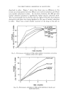
STATEMENT OF PROBLEM AND CRITICAL REVIEW OF PAST METHODS 67 cutaneous surface and the blood concentration of this substance is deter- mined at stated intervals, the amount of material which has entered the e determined from the calibration curve blood through the skin can b . . This method is most accurate when the rate at which the materrol reaches the blood stream is the same for the two methods of administration. Av. alysis of a single body tissue does not, of course, give information concerning total penetration it only shows the amount which has become localized in the tissue being analyzed (23). At times, this may be a very important weasurement if one is studying a specific, toxic reaction which mav follow percutaneous absorption. CONCLUDING REMARKS Loss of Penetrating Substance from the Surface If a known amount of a nonvolatile material which has been allowed to remain on the cutaneous surface for a definite period of time is quantita- tively removed from the surface and analysed, the difference between the amount applied and the amount removed may be thought to have pene- trated the skin (24). There are several pitfalls in this method. Quantita- tive removal is difficult. Some materials combine chemically with the constituents of the cornified epithelium and then cannot be easily separated from it although they actually have not penetrated into the skin any further than the stratum comeurn. The amount of most substances which penetrates is often so small that errors in analysis may be of the same order of magnitude as the amount absorbed, and therefore, one can never be sure that the difference between "before" and "after" determinations truly represents the amount of material which has penetrated. Now that radioactive substances can be prepared, it is possible to determine loss from the surface simply by determining the decrease in radioactivity (25). When some of the substance penetrates into the skin and is carried away by the blood stream, the radioactivity, as measured on the surface, decreases. Although other investigators have used this method successfully, it has not proved very satisfactory when used in our laboratory. Small movements of the experimental animal cause changes in radioactivity as measured by a Geiger tube held above the site of application. The amount of material which penetrates may be so small that the decrease in radioactivity is difficult to determine accurately. According to the title of this paper, I am supposed to have discussed "older" methods. There can be no clean cut distinction between "older" methods and "newer" methods. I have chosen to discuss some methods which have been in use over the years and have tried to point out some of their limitations. As knowledge has advanced, methods have been refined, and completely new techniques have become available. Ainsworth will
Purchased for the exclusive use of nofirst nolast (unknown)
From: SCC Media Library & Resource Center (library.scconline.org)