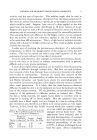
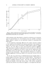
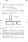
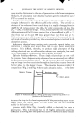
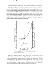
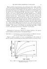
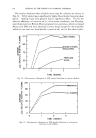
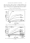
82 JOURNAL OF THE SOCIETY OF COSMETIC CHEMISTS because the epidermis presents a surface area 100 or 1000 times greater than the other two (2). Of course, under special conditions the sweat gland or sebaceous glands may be preferential pathways, e.g., during sweating of for lipide-soluble substances respectively. But for most conditions and substances the direct path through the epidermis is probably the impor- tant one. A general idea of the effect of cutaneous anatomy on permeation may be obtained from Table 1 in which are listed the relative values for rate of TABLE ]--PERMEABILITY OF VARIOUS LAYERS OF EXCISED HUMAN SKIN TO WATER (2, 3) Stratum Living Epidermis Corneum Barrier and Dermis Thickness (microns) 20-40 10-20 Permeability Rate J•/(t..4) 25 0.2-1.0 Permeability Coe•cient (J•.x)/(t..4) 500-1000 2-20 2000-4000 10 20,000-40,000 permeability of water outward through various anatomical zones of the skin (calculated from data by Blank (3) and Mali (2). Permeability co- efficient is a measure of the ease with which a substance passes through a zone. This concept has been developed further by Higuchi (4). Ob- viously, the barrier restricts water permeation 100 to 1000 fold. These observations hold also for other substances whose preferential route of penetration is the epidermis, but no data are yet available for the various zones encountered in sweat gland or sebaceous gland penetration. Let us now consider in more detail the biochemical and physiological aspects of the various anatomical zones as they affect permeability of the skin. Surface Film. The surface film is composed of sebum, sweat and des- quamating stratum corneum. Its chemical composition is complex and variable. The character and extent of its interaction with a penetrating agent are exceedingly difficult, if not impossible, to predict. Due to move- ment of the skin and sloughing of the horny layer, the surface film is dis- continuous and offers relatively little resistance to the penetratingmolecule. Stratum Corneum. On the other hand, the stratum corneBro has a greater effect on the penetrating substance. It may be quite thick as on the palms and soles. It is composed largely of keratin, a sulfhydryl contain- ing protein which adsorbs large amounts of water and other polar com- pounds. In addition, surface lipides, which may spread along the channel walls between cells, will retain lipide-soluble material. Thus, the stratum corneum acts like a sponge, becoming a reservoir for the penetrating agent and maintaining a maximum concentration gradient just above the bar- rier. However, if it binds the penerrant very strongly, it will actually hinder penetration. This is particularly true for ions and dyes of low molecular weight. As shown by autoradiography, ionized surfactants

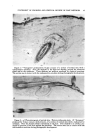
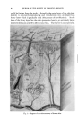
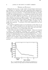
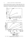
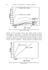
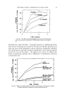
BIOLOGICAL FACTORS AFFECTING PERCUTANEOUS ABSORPTION 83 tagged with S a5 are bound by the horny layer and often do not penetrate beyond the orifice of the hair follicle (5). This adsorptive feature of the stratum comeurn serves a purpose, namely to bind noxious material until it is extruded into the external environment as the horny layer is shed. The water-binding capacity may affect penetration in another way: when the horny layer is well hydrated during sweating, hydrophilic cornl•ounds will reach the barrier more readily and lipophilic substances less •eadily. Barrier. The barrier zone has been isolated by Szakall (6) by adhesive tape stripping, but in sheets too small for use on diffusion chambers. It has been demonstrated histochemically by Stoughton (7) by means of dinitrofiuorobenzene which stains sulfhydryl groups in the barrier quite intensely. Just why sulfhydryl groups elsewhere in the stratum comeurn and living epidermis do not stain is not clear, but the technique is useful for visualizing the location of the barrier. The chemical structure of the barrier is unknown, but it must be quite similar to the living cells beneath and the dead horny cells above. The transition is abrupt, and one can only speculate about the nature of the forces which so suddenly cause the living epidermal cell to lose water and to bind its solid constituents so tightly. Electromicroscopy by Selby (8) of the junction between barrier-layer cells exhibits sturdy intercellular bridges and empty intercellular spaces, material having been lost during preparation of the tissue. Whether or not these spaces are artifacts or exist in life does not matter so much as what material is in the spaces. Certainly the intercellular regions which give rise to the spaces must be less tightly organized than the bridges and would be the avenue of penetration. It is known that molecules of greater than 200 or 300 molecular weight do not penetrate the intact barrier (9) yet the diameter of the pores in the material in the intercellular spaces in the barrier is much larger than the largest molecule penetrating. Thus, the restraining forcemust be the molec- ular interaction between penerrant and pore contents. If the substance has a high electrostatic charge, e.g., ions, the attraction is so great that no penetration occurs. If the substance has a water-lipide partition coefficient of about 1.0 it has the highest permeability the barrier then must have both polar and nonpolar groups in the pore contents. Living Epidermis. When the penetrating molecule reaches the living epidermis, its water solubility permits il to leave the barrier and continue on into the lower epidermis. Here, substances which are metabolized by cellular enzymes may stop their transit provided they can penetrate the epidermal cell walls but even these substances preferentially pass on to the dermis beneath because there is less resistance in the intercellular channels than in the epidermal cell walls. Dermis. At the junction between epidermis and dermis an electron- dense zone may be recognized in electron micrograms. But little resist-
Purchased for the exclusive use of nofirst nolast (unknown)
From: SCC Media Library & Resource Center (library.scconline.org)