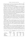
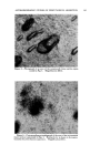
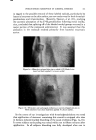
AUTORADIOGRAPHIC STUDIES ON PERCUTANEOUS ABSORPTION By B. R. C•oMa•* Presented September 23-24, 1959, Seminar, New York Ciu SUBSTANCES ABSORBED through the skin can be studied by chemically analyzing the blood, urine, internal organs or the expired air. However, the absorption of low concentrations of substances cannot be detected because of the limited sensitivity of the assay methods. To circumvent these difficulties, radioisotopes have been employed to demonstrate per- cutaneous absorption. Early investigations reported in the literature were made by analyzing body tissues and body fluids for the radioisotopes which had been applied to the surface of the skin (3-7). Development of the technique of autoradiography initially reported by Axelrod and Hamilton (1) has been shown to be valuable for determining the presence of tagged substances in histologically prepared skin sections. In reviewing the recent literature on autoradiography (2, 8-10), it was observed that many difficulties were involved in employing this technique. Because the photographic emulsion is sensitive to many forms of radiation contained in tissue exposed to radioisotopes, artifacts have been reported as true findings. It is important to note that tissue not exposed to radio- isotopes may cause the photographic emulsion to darken if improperly handled. Another difficulty has been the scattering of radiation from the radioactive tissue. Interpretation of the radiation located some distance away from the cellular structure is not always accurate. In most cases, autoradiographs of percutaneous absorption reported in the literature have been prepared from tissue cut perpendicular to the skin surface. The carrying of radioactive material from the surface of the skin to the underlying layers by the microtome knife can produce erroneous results of absorption. An improved autoradiographic method is presented which allows the detection of small quantities of radioactive substances in the skin resulting from topical application. This method avoids certain pitfalls inherent in older methods. The treated skin specimen is excised with a minimum of handling and it is kept frozen until processed. The histologi- * Lever Brothers Co., Edgewater, N.J. 138

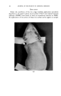
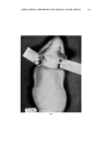
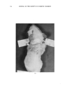
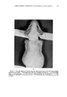
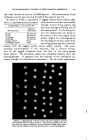
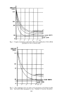
AUTORADIOGRAPHIC STUDIES ON PERCUTANEOUS ABSORPTION 139 cal sections are made by cutting the skin parallel to the surface. By first sectioning the distal dermal layer of tissue little or no radioactive contamination is imparted to the microtome knife or to each serial section. The method given here provides a means of obtaining information on the rate of percutaneous absorption and also sheds light on the routes of pene- tration. Because of our interest in learning about the absorption of surface active agents by the skin, sodium lauryl sulfate labeled with S a5 was chosen to be studied by means of the new technique. In addition, the absorption of nickel chloride labeled with Ni 6a was investigated. The studies were conducted with the laboratory rat and the guinea pig serving as the source of skin specimens. Experimental Methods The hair on the dorsal area of the animal was removed by means of an electrical clipper without damage to the skin. The animal was im- mobilized on a board to prevent body movement and to prevent spreading of the radioactive material. The isotope solution was applied by means of a micropipette and was permitted to dry on the skin surface. Rinsing of the skin before or after treatment was not employed. The studies were conducted in a laboratory maintained at 24øC., with a relative hu- midity of 60 per cent. After the desired contact time the animal was sacrificed by exposure to ethyl ether. The dorsal skin was removed from the animal immediately and placed on a wooden block with the epidermal side facing down. By first inserting a skin biopsy punch (6 min. diameter) through the subcutis layer, the specimen was removed without directly contacting the applied radioactive material. The tissue was placed at -4øC., for immediate freezing. The frozen specimen was transferred to the freezing microtome with the epidermal side facing down. Attachment of the specimen to the freezing block was made with a small single drop of water rapidly frozen with carbon dioxide. No additional water was placed on the speci- men before sectioning. Manipulation of the tissue in this manner elimi- nated leaching of radioactive material from the tissue it also prevented contamination of the microtome. The entire tissue specimen was cut parallel-to-the-surface beginning with the subcurls layer. Each tissue section of 25 microns was removed from the microtome knife with a fine glass rod and arranged sequentially on a glass plate 12.5 cm. square. Care was taken to unfold each section so that it contacted the glass uniformly. The glass plate containing the serial sections was pressed against a sheet of Eastman Kodak medical x-ray film and was sealed in a light-proof container. Incubation was made at 4øC., for a period of time sufficient
Purchased for the exclusive use of nofirst nolast (unknown)
From: SCC Media Library & Resource Center (library.scconline.org)