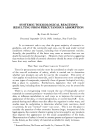
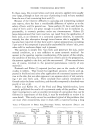
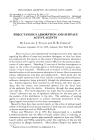
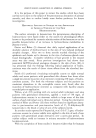
140 JOURNAL OF THE SOCIETY OF COSMETIC CHEMISTS to produce a complete reaction on the film. Depending on the activity of the isotope solution and quantity applied, the incubation time varied from three to four weeks. The x-ray film was developed with Kodak rapid x-ray developer for six minutes, and then placed in Kodak x-ray fixer and replenisher for ten minutes. Development of the film showed the relative intensity of radiation in the individual tissue sections. By determining the number of serial sections which contained sufficient radiation to promote darkening of the film, calculation of the depth of absorption through the skin was made. STUDY A The following experiments are presented as examples to illustrate the utility of the autoradiographic procedure described. In the first study, normal guinea pigs were employed. On the dorsal shaven area of each animal, one treatment of fifty lambdas of Cj2H25S3504Na was made. The radioactive material was permitted to remain on the living skin for sixty minutes. No rinsing after treatment was employed. The animal was sacrificed immediately by exposure to ethyl ether. The autoradiograph of the treated living skin was prepared as described in the preceding section. The remaining dorsal skin from the sacrificed animal was removed four hours after death and was considered to be nonliving. The subcutis layer was excised carefully by means of a scalpel. The non-living skin was divided into four individual squares of 2 cm. Two of the skins were secured over the top of a stainless steel chamber (2 cm. diameter X 0.6 cm. high) containing four ml. of distilled water which was in contact with the dermal layer. Fifty lambdas of the tagged surfactant were placed on the skin surface. Diffusion of the radioisotope through the skin was determined by analysis of the underlying water after thirty minutes, one hour, two hours and twenty-four hours. Aliquots of the water were placed on a glass plate and evaporated under an infrared lamp before being placed in contact with the x-ray film. The presence of radioactive material in the sample was detected by the production of dark zones on the developed x-ray film. The other two nonliving skins were attached to a wooden board without being exposed to the water chamber, and one treatment of fifty lambdas of tagged surfactant was applied to the skin surface. After sixty minutes a specimen of the treated site was taken by means of the skin biopsy punch and the autoradiograph of the tissue was prepared. In addition, prior to and after death, skin specimens were removed from each animal by means of the skin biopsy punch to represent untreated tissue. Autoradiographs of the living and nonliving control skins were prepared in the same manner as the treated skins. The radioactive solution of tagged sodium lauryl sulfate employed in

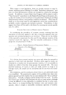
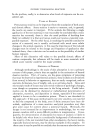
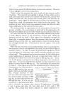
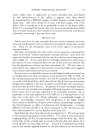
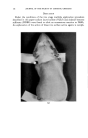
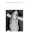
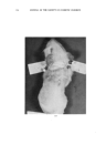
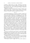
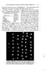
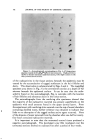
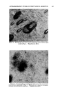
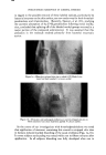
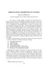
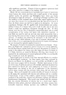
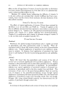
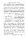
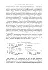
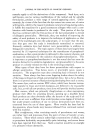
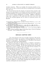
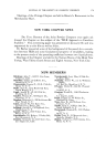
AUTORADIOGRAPHIC STUDIES ON PERCUTANEOUS ABSORPTION 141 this study showed an activity of 63,000 dps/ml. The concentration of the surfactant was 0.5 per cent and the pH of the solution was 7.2. As shown in Table 1, absorption of tagged sodium lauryl sulfate after TABLE 1--AuToRADIOGP,•PHIO DETERMINA- TION OF TIlE PERCUTANEOUS ABSORPTION OF C12H2•Sa•'O4NA IN TIlE GUINEA Px• Depth of Skin Specimen Absorption Living 750 microns Living 800 microns Nonliving 200 microns Nonliving 250 microns Living, control None Nonliving, control None sixty minutes occurred more readily through normal living guinea pig skin than through nonliving skin of the same animal. In the living tis- sue, the radioisotope was found to be present in the lower region of the dermis. Figure 1 is a photograph of the histological sections made from one living guinea pig skin specimen treated with the tagged sodium lauryl sulfate solution. The corre- sponding autoradiograph of this specimen, Fig. 2, showed absorp- tion of the tagged surfactant from the first to the thirty-second serial section. The absorption depth was calculated directly from the autoradiographs. It appeared that the majority of the absorption oc- curred through the pilosebaceous apparatus. The pin-point appearance 6 .! I Figure 1.--Photograph of living guinea pig skin specimen treated with C•2H2•Sa•O4Na for sixty minutes. Sections cut parallel to the sur- face with the external epidermal section located in lower right corner. Other sections follow in sequence from bottom to top of photograph.
Purchased for the exclusive use of nofirst nolast (unknown)
From: SCC Media Library & Resource Center (library.scconline.org)