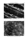
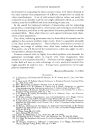
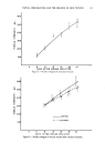
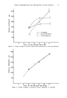
490 JOURNAL OF THE SOCIETY OF COSMETIC CHEMISTS measured volume would be 160/z a. The minimum measurable amount of absorbing substance in this volume then is approximately 2 X 10 -•ø Mole. With a molecular weight of around 250, which again is an average value for a typical dye molecule, this sets the minimum detectable amount of such a dye at 5 X 10 -•4 g. Certain precautions have to be taken. For instance, it is advisable to use an unstained but otherwise identical specimen to measure I0, in order to compensate for light losses due to light scattering. If the absorbing material is not distributed homogeneously but in the form of granules, one has to use the two-wavelength method (3). Microspectrophotometry has a number of applications in the cosmetic industry, especially so in histochemical work or in penetration studies. Some materials reveal more information in reflected light than through their absorption spectrum, as shown in Figs. 3 and 4. Figure 3 shows the absorption spectrum of human hair, covering the range from bleached, almost white hair to dark black. The absorption spectra in the visible range are not very informative. Figure 4, however, shows the spectral reflectivity. The technique is the same, in principle, as that employed in transmitted light, but the condenser of the microscope is replaced with a vertical illuminator. Since hair is highly birefringent, the same precau- tions have to be employed that are also taken in measurements of the re- flectivity of ores and minerals (4, 5). Similarly, not only the relative spectral reflectivity can be measured, but also, in suitable cases, changes in reflectivity, e.g., after treatment of hair with cosmetic preparations. Again, once a measurable parameter is found, valid comparisons become possible. PHASE {•JONTRAST MICROSCOPY Many specimens, however, do not contain light absorbing structures. Their detail consists of structures with a refractive index different from that of their surroundings. In a normal brightfield microscope, such structures do not appear with any appreciable contrast. Such phase de- tail can be made visible by phase contrast (6, 7). Differences in optical path are by this process converted into differences of light intensity. Inherent in the process, however, is the occurrence of light or dark seams wherever steep gradients of refractive index occur. These artifacts are called halos, and due to their presence the intensity distribution of a phase contrast image cannot be used to determine quanti- tatively the optical path differences in the specimen. For qualitative observation, however, phase contrast is ideal in the study of pure phase objects, in the examination of washing agents, the

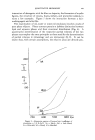
QUANTITATIVE MICROSCOPY 491 interaction of detergents with fat films or deposits, the formation of myelin figures, the structure of creams, foams, lathers, and protective colloids, to name a few examples. Figure 5 show• the interaction between a hair- washing agent and a fat film. The examination of oil, water or water/oil emulsions involves study of pure phase objects. Phase contrast permits a definite distinction between lipid and aqueous phases and their structural distribution (Fig. 6). A quantitative determination of the respective partial volumes of the two phases may employ the same principles as those used for the determination of partial volumes in mineralogy and ore microscopy (8, 9). It can be shown that, with certain assumptions, the relative areas are directly pro- 2OO I d 700 600 5 O0 400 m•zz Figure &--Absorption spectra of human hair (coefficient of absorption = 1/d In Io/I. bk = black dbd = dark blond dr = titian red bd == blond bl --- bleached.
Purchased for the exclusive use of nofirst nolast (unknown)
From: SCC Media Library & Resource Center (library.scconline.org)