502 JOURNAL OF THE SOCIETY OF COSMETIC CHEMISTS To convert this value into differences of concentration of a substance, one has to know the specific refractive increment, i.e., the increase of the refractive index of a solution for every 1% increase in concentration. For most biological materials, such as proteins, carbohydrates or lipids, this value is 1.85 X 10 -3 (13). Using this value, it follows that an in- crease in concentration of about 0.2% could be detected. In order to ob- tain practical numerical values for the specific refractive increment the concentration is expressed in g./100 cm. 3. The volumes of biological cells or cell components are very small. A volume of 15• 3 shall be assumed. Since a change of 200 rag. in 100 cm. 3 is detectable, in a volume of 15• 3 this amounts to only 3 X 10 -ng. In this application, the interference microscope thus becomes a highly sensitive optical balance. Such a highly sensitive method is not only applicable to biological prob- lems but also offers opportunities in the study of dissolving rates, of the effect of protective colloids added to soaps and lathers, and interactions between detergents and films of a fatty nature. Interference microscopical methods thus offer a possibility to measure quantitatively those effects of a cosmetic treatment or preparation and of colloid chemical processes which produce a change in optical path. For practical purposes, measurements can detect changes of optical path from around 1 min. to 20 A., a range of approximately six orders of magnitude. ANALYSIS IN POLARIZED LIGHT One of the most revealing microscopic methods is the examination of microscopic structures in polarized light. Numerous materials are either crystalline, as, for example, many organic substances, or they have sub- microscopic crystalline regions, for instance, gels (23, 24). Such structural anisometry leads to optical anisotropy. The anisotropy characteristics of such specimens can very accurately be measured in a polarizing microscope by analyzing the state of polarization of light which has passed through them. Even the molecular arrangements in the structures of these mate- rials can be derived from such studies. Analysis in polarized light can lead to highly interesting information on the orientation of submicroscopic elements, micelles and gel components in anisotropic materials. On the other hand, polarized light can also serve as a highly sensitive method of detection. In fact, in some specialized polarizing microscopes with rectified optics, optic path differences of as little as 0.1 A. have been detected (25). But even though the sensitivity of standard polarizing microscopes will not go quite as high, it is more than adequate for most purposes. The full measuring range of a 1/30 wavelength mica compensa- tor covers 170 A., and its lower sensitivity limit is of the order of magnitude

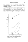
QUANTITATIVE MICROSCOPY 503 of a few Angstrom units. What then does an assumed sensitivity of 5 A. mean for a practical application in cosmetics research ? One practical example is the study of hair. Hair as such is rather highly birefringent, which makes it difficult to measure small changes against the fairly nonuniform background. Nevertheless, it may be interesting to calculate, as a numerical example, how thin a coating of birefringent mate- rial on the surface of hair can be measured in a polarizing microscope, or how much birefringent material would have to penetrate into the interior of a hair to change its anisotropy. The total birefringence of hair would, by the way, already be affected by any penetration (even of isotropic materials) due to a change of form birefringence. For the purpose of this calculation, a polymeric hair conditioning agent shall be assumed to coat the hair. Assuming a value for the birefringence of oriented polymer material of 0.02, we can then calculate the minimum thickness necessary for such a coating to be measurable. This follows from the equation F = t (n•. -- n•), in which I' represents the retardation or optic path difference in m•, t the geometric thickness, also expressed in m•, and n•. - n• the birefringence. F•in was assumed as 5 A., or 0.5 m•, n•. - n• as 0.02. The minimum value for t then becomes 25 m•, or 250 A. The total birefringence of a structure like a human hair is the result of the contributions of the different histological components, such as the medul- lary and the cortical cells, and the total birefringence of each of these com- ponents again is the sum of their textural and their intrinsic birefringence. The intrinsic birefringence is a material constant the textural birefringence, also called form birefringence, is a function of the orientation of the in- trinsically birefringent elements and of their partial volumes, compared to the partial volume of the unoriented matrix. Any mechanical influence, such as mechanical stress which leads to elastic or plastic deformation, will affect form birefringence (23), the changes of which can be used as sensitive indicators. Swelling and thermal treatment will not only affect form birefringence but also intrinsic birefringence. FLUORESCENCE MICROSCOPY Fluorescence microscopy in its various applications combines highest sensitivity of detection with extreme specificity. It utilizes the fact that many substances become self-luminous when irradiated with light of short wavelengths, i.e., with light from the energy-rich blue and ultraviolet range of the spectrum. This irradiating or "exciting light" stimulates fluo- rescence in such substances. The fluorescent light always has a longer wavelength than the exciting light. Almost any microscope can be adapted for fluorescence microscopy. Some applications require hardly any, others demand more elaborate
Purchased for the exclusive use of nofirst nolast (unknown)
From: SCC Media Library & Resource Center (library.scconline.org)