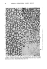
504 JOURNAL OF THE SOCIETY OF COSMETIC CHEMISTS accessories. First, a light source is needed which yields a powerful flux of short-wavelength light. Maximum pressure mercury arcs are necessary for all work involving immuno-fluorescence (26, 27) and for work carried out under oil immersion with other staining techniques. For medium and low power work, where fluorochromes like Acridine Orange are employed, adequate excitation can often be obtained from a high intensity incandes- cent lamp. All such light sources do, of course, not only emit the desirable short wavelengths, but also light of longer wavelengths, which would com- pletely mask any fluorescence. For this reason, all exciting light is filtered through a set of exciter filters, which transmit only the short wavelength range of the spectrum and completely absorb light of longer wavelengths. The exciting light is then concentrated on the specimen by a microscope condenser. In immuno-fluorescence this is normally a dark field condenser to give images of very high contrast, but in most other work a regular microscope condenser with all diaphragms wide open is sufficient. Fluo- rescence is stimulated in the specimen and both the remaining exciting light and the stimulated fluorescence enter the objective. To remove any of the remaining short wavelength exciting light, a barrier filter is mounted above the objective. It has a transmission curve complementary to that of the exciting filters and absorbs all light of shorter wavelengths than that of the fluorescence. The specimen, therefore, appears in brilliant luminosity against a dark background. Many organic substances have the intrinsic property to fluoresce (28) and can be traced directly in a microscopic preparation. Others can be stained selectively with fluorescing dyes, so-called fluorochromes this fluorescence is called secondary fluorescence. Finally, a fluorescing molecule can be used to tag an otherwise nonfluorescing complex, such as an antigen or anti- body. It is in this last procedure that the high sensitivity of detection inherent in fluorescence techniques can be combined with the extreme specificity of serological methods. This technique is known as the fluores- cent antibody technique, or immuno-fluorescence. It may be that the remarkable success and the exciting potential of this latter technique has diverted the interest in the field of fluorescence microscopy from those simpler staining techniques which can be used so successfully in routine work. Among the many fiuorochromes which are available for fluorescence staining, Acridine Orange has found some interesting applications. For example, under certain conditions, it permits differentiation between living and dead protoplasm (29). Acridine Orange, or tetramethyl-diamino acridine, is a basic dye. In the alkaline pH-range it is present as an un- charged base molecule, which selectively stains lipid phases with a dark, saturated green fluorescence. In the weakly alkaline, neutral or acid range, however, Acridine Orange is present in solution in the form of a univalent

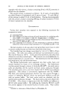
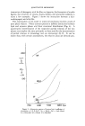
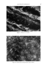
QUANTITATIVE MICROSCOPY 505 cation. In dilute solutions (10 -6 M) the cation exists as a monomer and fluoresces bright green. With increasing concentration dimerization sets in. The dimer has an emission spectrum with its maximum in the red spectral region, so that the fluorescence color indicates the dye concentra- tion. This so-called concentration effect has successfully (29) been em- ployed to distinguish between living and dead protoplasm. This applica- tion permits rapid testing of the effectiveness of antibacterial agents in skin preparations, deodorants or toothpastes by bacterial counts and replaces, when applicable, the time-consuming growing of colonies. To obtain re- producible results it is necessary to control the pH carefully with buffers, to use excess dye solution, and to stay within a certain pH-range. Also, it is necessary to check whether the tested preparations contain fluorescence quenching agents and whether the bacteria under examination show the typical green-red transition (Fig. 15). The uncharged molecules of several basic dyes accumulate readily in lipid phases. Addition of a fluorochrome to fatty substances allows determina- tion of the uniformity of very thin films of these substances, or their pres- ence, or their penetration. One of the great advantages of fluorescence techniques is, as has been mentioned before, the very high sensitivity of detection. One can clearly recognize a particle as stained when the concentration of dye in it is as low as 1:10,000. For particles smaller than 1•, which show up very clearly Figure 15.--Mucosa cells with bacteria stained with Acridine Orange (320X).
Purchased for the exclusive use of nofirst nolast (unknown)
From: SCC Media Library & Resource Center (library.scconline.org)