510 JOURNAL Oh' THE SOCIETY OF COSMETIC CHEMISTS restored to its normal state. There are shifts in the mucopolysaccharides, glycoproteins, amino acids and RNA concentrations (1-7). Cellular migrations and multiplications occur to replace lost tissue (8), and complex biophysicochemical reactions result in the formation of collagen fibers which help to restore tissue strength (3, 5, 9). The effects of a wide variety of substances, administered both system- ically and topically, on the reparative processes have also been studied. Nutrients, hormones, antimicrobial agents, botanicals and an array of pharmacologically active compounds have been evaluated. Among these, cortisone derivatives, Acr['H, thyroxine, hyaluronidase, reserpine and 5- hydroxytryptamine have been reported to retard the healing of wounds (10-19). Promotion of healing has been ascribed to glycosides of Centella asiafica, extracts of,4/oe vera, a]lantoin, protein hydrolysates, nitrofurans, pantothenates and hyaluronic acid (20-30). Gross and histological observations, changes in the concentration of various tissue constituents, and the strength of the healing wound are the criteria most often used to determine healing rates and the effects of treat- ment upon them. As has been mentioned, many biological and biochemical mechanisms come into play during healing and may be considered as forces directed toward returning traumatized tissue to a normal condition. The strength of the healing wound is a general measure of the progress toward normalcy and may be thought of as the biological resultant of the various forces acting in and on the affected area. It is a comparatively simple measurement and represents a direct approach to the problem (9, 19,31,32). Two general methods of skin wound tensiometry have been described and used. Prudden and co-workers (33-41), by measuring the pressure, applied from within, required to disrupt abdominal incisions in the rat, have reported that the application of ground, acid-pepsin digested, bovine tracheal cartilage stimulated healing. The other technique consists of determining the force (usually as measured by weight) required to cause the separation of the edges of a standard segment of healing skin wound. This wound-strip method was used in the studies reported here. Standardization of the method of injury has also been approached in a variety of ways. Burns, blows and cuts have all been used in the reports cited. A surgical incision of closely controlled length and depth was used in our work, since it could be reproduced with acceptable precision. MATERIAlrS AND METHODS Test Atnimals CF-1 male mice, 30-40 days old, and weighing 18 to 25 g. were used in all the experiments.

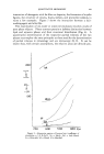
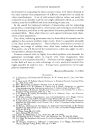
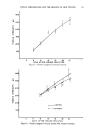
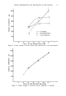
TOPICAL PREPARATIONS AND THE HEALING OF SKIN WOUNDS 511 lP'ounding Procedures On the day prior to inflicting the wounds, the hair on the backs of the animals was removed with an electric clipper. With the animals under ether anesthesia, an incision approximately 25 mm. in length was made along the midline of the back, starting at the shoulders and proceeding caudally. In order to cut only the skin and not involve underlying tissue, an initial small slit was made in the skin with a sharp, fine pointed scissors. The skin was then raised by grasping it with forceps at a point just an- terior to the slit and a scapel blade, cutting edge upward, was inserted into the opening and drawn through the skin to a point about 25 min. from the point of origin. The edges of the wound were approximated and then closed with five equally spaced interrupted sutures (40 gauge stainless steel wire). Treatment was begun immediately. On the sixth day after the wound was inflicted, the sutures were removed. Tensile Strength Measurements The mice were sacrificed and hair that had regrown was carefully clipped with a small scissors. A wide section of skin containing the healing wound was excised. Care was taken to include only the skin and closely adhering subcutaneous tissue. The skin section was then placed on a plate-glass block that was moistened with normal saline. This permitted the skin to assume what might be termed a standard dimension. A strip of skin along the posterior (caudal) end of the wound, perpendicular to the line of the wound, was trimmed off and discarded. Then a strip 8 mm. wide was cut off and used for the tensile strength measurements. The next 2-3 mm. section of the wound was removed and fixed in formalin for histological examination. Another 8 min. segment was then obtained from the re- maining (anterior) portion of the wound, for a second tensile strength de- termination. A modification of the Charney, Williamson, Bernhard (42) technique was used to determine the tensile strength of the healing wound segments. One end of the skin section was mounted in a clamp, and a lightweight basket lined with a polyethylene bag was attached to the other end. With the skin segment in a vertical position, weight in the form of dry, free flowing sand at a rate of 7 g./sec. was added to the basket until rupture of the wound occurred. The results are expressed as grams or weight required to separate the edges of the healing wound. Averages for each group were calculated and include the values obtained with both segments of wound from each mouse. Four series of tests were conducted. The first was for the purpose of checking the basic method and to construct a time rs. wound tensile strength curve, with the animals under control conditions, i.e., no treat- ments applied to the wounds. Following this the experiment was re-
Purchased for the exclusive use of nofirst nolast (unknown)
From: SCC Media Library & Resource Center (library.scconline.org)