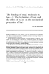
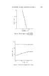
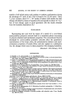
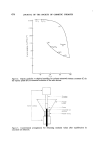
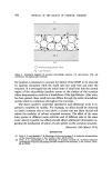
484 JOURNAL OF THE SOCIETY OF COSMETIC CHEMISTS The effective performance of this 'barrier function' appears to depend on the chemical composition of the stratum corneum. Chemical analysis has shown that this layer is rich in 'solid' matter which consists principally of keratin fibrils and lipid. These components are derived from the stratum spinosum, the layer of living cells immediately beneath the stratum lucidum (33, 46). The investigations of Matolsty, Downes and Sweeney (47) have shown that removal of the lipid or the protein of the stratum corneum results in an increased permeability to water indicating that both are essential to maintain intact the barrier property of this layer. As one would expect, the thicker the stratum corneum, the more im- permeable is the barrier. Marzulli (29), Blank and Scheuplein (33) and McCreesh (48) have shown that the rates of penetration of a wide variety of liquids applied to the epidermal surface in vitro were inversely related to the thickness of the epidermis, so that the soles and palms are much less per- meable than the skin in other parts of the body. It would appear, however, that this rule does not apply in vivo. In a study of the comparative rates of absorption of parathion, malathion and carbaryl from a number of anato- mic sites, the x4C-labelled compounds were absorbed with equal facility from the palm or the hand or from the forearm. The abdomen and dorsum of the hand had twice the penetration of the forearm, whereas follicle-rich sites, including the scalp, angle of the jaw, the area behind the ear and the forehead, had a four-fold greater penetration (49), indicating that the skin appendages may constitute an important pathway for absorption. The role of the appendages The barrier formed by the stratum corneum is broken by the ducts of numerous glands and hair follicles, which, as indicated above, form an additional route by which substances applied to the skin surface might gain access to the circulation. In the human, the density of sweat ducts is 210 and 220 cm -•' in abdomen and forearm respectively (50), while that of the hair follicles is 40 and 100 in these same areas. This density is, however, variable--both between different anatomical areas and between species. The presence of this additional route of percutaneous absorption has been verified by a number of experiments. Applying dyestuffs on the skin in vivo Rein (51) showed that the vicinity of follicles is stained more intensely than the rest of the epithelium. He also found that perifollicular staining does not occur around follicles which have been occluded by waxy plugs. These observations were supported by the work of MacKee, Sulzberger,

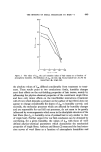
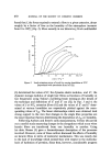
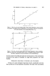
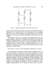
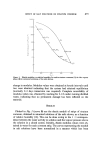
PERCUTANEOUS ABSORPTION 485 Hermann and Baer (52) who studied the penetration of heavy-metal com- pounds, sulphonamides and dyes through the skin of guinea-pigs and men, using histochemical techniques for the identification of these compounds. Further evidence has been provided from experiments using isotope- labelled compounds: for example, in studies of the passage of labelled para- thion through the skin of a variety of species, including man, a high concen- tration of labelled material was found in the appendages (53) suggesting a preferential absorption via this route. In an appraisal of these autoradio- graphic results Fredriksson (54) suggested that they could equally well be regarded as an affinity of the test substance for certain structures. The same critical comment could be applied to observations on the localization of coloured compounds or on the histochemical demonstration of other compounds. Percutaneous passage by way of skin appendages was elegantly demonstrated by Van Kootan and Mali (55). Employing in vitro techniques, these authors mounted a freshly obtained piece of cadaver skin in a Perspex chamber and perfused one side of it with potassium ferrocyanide, and the other side with ammonium ferrisulphate. The solutions diffused through the epidermal barrier and formed a blue precipitate (Turnbull blue) at the site where they met. Under these conditions it was shown that a precipitate of Turnbull blue was formed at the epidermal barrier and within the skin appendages, especially sweat glands. The contribution of sweat glands and of hair follicles to the percutaneous absorption of test substances has been investigated by a number of workers. Substances that are absorbed via the appendages must first pass through the orifices at the skin surface. According to Scheuplein (50) this process takes place by diffusion since 'hydrodynamic flow is excluded'. Taking into account the differences in the diffusion constants of the keratinized epider- mis and the cells lining the ducts of the appendages, this author calculated that the route via the glands is important for a brief period immediately after the application of the test substance. During this period, absorption is greater through the appendages than through the matrix of the stratum corneum. Later a 'steady state' is established during which the bulk of diffusion appears to be no longer intra-appendageal but occurs through the matrix of the stratum corneum (50). In his studies, Scheuplein (50) considered only water and water-soluble substances and precluded the effect of solvents such as chloroform and methanol since these destroy the stratum corneum and as a consequence alter considerably the dynamics of absorption. In the case of lipid-soluble substances which do not affect the integrity of the stratum corneum to any appreciable extent, absorption via the pilosebaceous
Purchased for the exclusive use of nofirst nolast (unknown)
From: SCC Media Library & Resource Center (library.scconline.org)