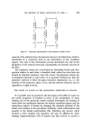
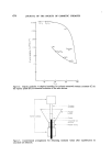
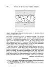
486 JOURNAL OF THE SOCIETY OF COSMETIC CHEMISTS apparatus may be considerable, due to its lipid-rich sebaceous secretion (50). The results obtained by Maibach et al (49) in their study of the percutaneous absorption of the lipid-soluble insecticides, parathion, malathion and carbaryl, mentioned in an earlier section, give considerable support to this hypothesis. Further light on the relative rates of absorption via the skin appendages and the stratum corneum was provided by Wahlberg (22, 56). He used a solution of •'•'NaC1 and applied it to the shaved abdominal skin, rich in hair follicles and to the non-hairy skin behind the ear in guinea-pigs. From in vivo and in vitro experiments lasting several hours he showed that the percutaneous absorption from the abdominal skin was approximately 20•o greater than that from the non-hairy skin and attributed this difference to the amount absorbed from the greater number of hair follicles in the abdominal skin. This conclusion was supported by a second experiment in which he used the same technique but substituted HgCI•. for the sodium chloride. No difference in the absorption rates of Hg was observed between the hairy and non-hairy skin. This was due to the occlusion of the hair follicles and their glands by the protein precipitated by the mercury. These results suggest that even during the 'steady state', absorption via the hair follicles forms an appreciable fraction of the total amount absorbed. It would seem reasonable to assume that the sweat glands, if present, con- tribute an amount at least equal to that of hair follicles to percutaneous absorption. While the hair follicle may complicate measurements of the amount absorbed, the hair keratin may interfere with measurements of the amount of the compound retained in the skin (57). In the design of experiments of this sort, it is important to remember that considerable regrowth of hair may occur, especially in young animals, within a few days of shaving. METHODS FOR TESTING Efficiency of the cutaneous barrier Of the procedures that have been employed for measuring the barrier properties of the skin, the one most frequently used is the water diffusion test, carried out under controlled humidity conditions in vitro (58, 59). A cylindrical aluminium cell containing water is separated from the atmo- sphere by a piece of skin which serves as a membrane. Water loss through skin after an appropriate equilibration time is expressed as mg cm

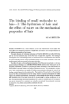
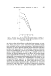
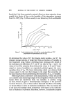
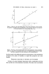
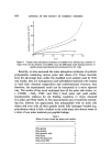
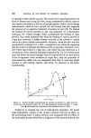
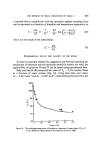
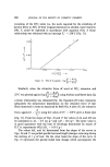
PERCUTANEOUS ABSORPTION 487 The permeability coefficient can be calculated if the water vapour gradient across the membrane is known. By appropriate experimental manipulations a comparison between the water loss through intact skin and through its ß component layers can be carried out. Studies of this sort have shown no change in the rate of water loss whether one uses whole skin, a preparation consisting of epidermis and stratum corneum or stratum corneum on its own. Vinson (60) conducted a comparative in vitro study of the rate of diffu- sion of water through normal skin from neonatal and adult rat, from the adult guinea-pig and swine, and from male and female adult human sub- jects. The skin from neonatal rat was the least permeable exhibiting the lowest diffusion values (0.15 mg cm -• h-•). The diffusion values for adult rats and guinea pig skin and for specimens from the back or abdomen of adult swine were about four to five times that of the skin from neonatal rats. Values for human skin varied between these two extremes. Specimens from the female thigh gave diffusion values close to those of the neonatal rat. Others taken from the calf or from the abdomen in the male gave values close to those derived from adult swine The skin from the sole of the foot, taken from an adult female gave values of 2.1 mg cm A simpler method for assessing the barrier properties of the stratum corneum is the measurement of its electrical conductivity. In the intact skin electrical conductivity is of the order of 1 I•A V -• and it is increased considerably after skin damage by abrasion or chemical action (60). Thus, immersion of guinea-pig skin in vivo at 70øC for 45 s, increased the con- ductivity from the control value of 0.04-0.9 ira V -• to 2.2 ira V-L The increased rate of conductivity correlated with histological damage and with increased water diffusion over the range 0-10 I•A V 4. Application of di- methyl sulphoxide (DMSO) in concentrations of 20•o or higher decrease considerably the electrical resistance of the skin (61). At such concentrations DMSO damages the keratin layer (62). A simple apparatus for electrical conductivity measurements has been described by Blank and Finesinger (63) and can be readily constructed. In practice, measurement of the effectiveness of the cutaneous barrier does not form part of the routine tests for measurements of percutaneous absorption. The water diffusion test is too elaborate to be recommended for inclusion in tests of this sort. The electrical conductivity test on the other hand is simple and could be readily carried out. It could be useful as a screening procedure where enhancement of percutaneous absorption is suspected to be due to damage to the keratin layer or to an increase in its
Purchased for the exclusive use of nofirst nolast (unknown)
From: SCC Media Library & Resource Center (library.scconline.org)