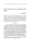
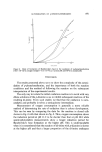
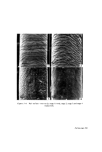
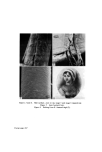
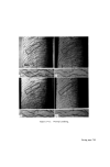
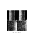
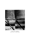
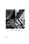
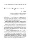
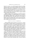
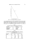
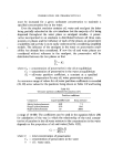
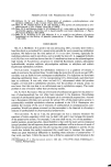
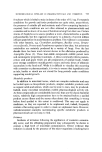
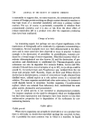
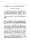
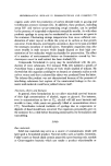
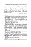
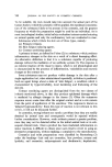
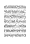
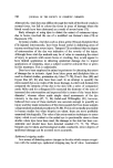
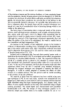
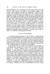
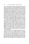
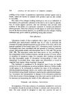
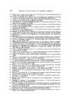
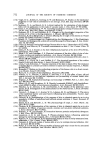
'• ,•5•'-•%.:.: . • . •:?-- •...--• . .?• • '• •, • • . '.. ...... .../.• _.•:.• '• ..... .. .... •. •. :.-•. . :• • .-'• '?.-"-z • :•,• ::: .• •. •. '• '-.. •'• - •.: • ?.' .... ,... :• • •. '•, •-. : : • •. : ß • '-•. .•.:,. z.: •. •.•: :-. '• ' . •. .... -..-. ". :• • ' • • . • • • •. '• . '• r .. • • • • % .... .. '•' .. ½•,, • • •. •. ..:•/-'-b'() %• '•-• • • •...' [: ,J• '• .• ... - • .•.- •. ..- • •. ........ •'• :?•¾: • ..... ?.• .... 2•-•'":•:• .%,.... •,• •e• .... ß ...... Figures 21 and 22. Polymer treatment. F/gures 23 and 24. Cuticle debris, surface and section respectively. Facing page 701

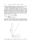
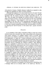
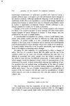
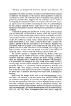
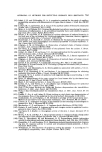
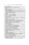
CHANGES IN SURFACE OF HAIR DUE TO COSMETIC TREATMENT 701 Finally, one of the clearest demonstrations of our present techniques has been in the examination of the split ends of naturally weathered human hair after the application of a polymer. The pairs of micrographs of Figs. 19-22 show that the disorganized cortical fibrils of the split ends are neatly replaced in the body of the hair fibre, restoring it to its original gross configuration. EXAMINATION OF THE FRAGMENTS BREAKING OFF HAIR AFTER COMBING AND MECHANICAL AGITATION The previous studies of hair surfaces have demonstrated that the scale edges of human hair break off with mechanical atrophy and it was therefore pertinent to examine the nature of the fragments released from the hair. The root end of a switch of untreated hair was thoroughly washed in distilled water with many rinses. After drying, one part of the switch was cut into pieces of 2 cm length, again washed thorougly with distilled water and then thoroughly shaken with distilled water in a flask on a laboratory shaker. Another part of the switch was combed (dry) with 2 000 strokes of a nylon comb and then rinsed in a small volume of water. The remainder of the switch was combed wet with 200 strokes of a nylon comb and this too was rinsed in a small volume of water. In each case the water was turbid and was therefore centrifuged. The various sediments were prepared for examination in the scanning electron microscope and also some embedded in epoxy resin, sectioned and stained for examination by transmission electron microscopy. Under the scanning electron microscope all the residues were seen to be composed of tiny platelets (Fig. 23) up to 5 gm in diameter and 0.3 gm thick. These are evidently fragments of the hair cuticle. In section (Fig. 24) the pieces are nearly all of one scale thickness and from their structure it would appear that they have been released mainly by cleavage along the cuticle cell membranes. A few of the pieces had also been released by cleavage through the endocuticle, a cystine-poor layer which constitutes one of the two major lamina which make up each cuticle cell (4). DISCUSSION Our experiments have shown that with treatments such as bleaching and perming and even with combing, the scale margins of human hair slowly chip away. The changes are relatively minor but nevertheless even with combing we can expect, and in fact do see, the gradual loss of the hair cuticle with time (i.e. from root to tip).
Purchased for the exclusive use of nofirst nolast (unknown)
From: SCC Media Library & Resource Center (library.scconline.org)