APPRAISAL OF METHODS FOR DETECTING PRIMARY SKIN IRRITANTS 747 effects, Schwartz and Peck (59) suggested that yet a further precaution might be taken, namely, that the product should be distributed to between 5 000 and 10 000 people. If no adverse reactions are reported, the product could be deemed safe for public use. The outcome of human patch testing may be influenced by variations in the sensitivity of the individual subjects and environmental factors. Bjornberg (60) reviewed the various human factors which could influence the response of human skin to primary irritants. He found that females were more sensitive to irritants than males, and white people more than negroes. In another study, Caucasians were found to give very consistent results in patch tests (11). It is likely that the variations in response to sex or race are due to inherent differences in the morphology of the skin, its biochemical constitution (61, 62), the rigidity of the skin at the test site and the degree of muscle action beneath it (23). This explanation may also account for the greater sensitivity of the skin in one area of the body than in another (24, 63). For example, the skin of the eyelids, which is devoid of hair and has only a thin layer of keratin (64), is considerably more sensitive than the skin on the soles of the feet, where it is thick and has a substantial layer of keratin. The sensitivity of the skin is dependent also upon the health of the individual at the time of testing. Skin which is subject to psoriasis is considerably more sensitive to irritants than normal skin (55, 65). The environmental influence on the outcome of human patch testing is such that the cutaneous response to irritants is greater in winter than in summer (4, 66, 67). In summer, when the skin is well hydrated as a result of sweat and sebaceous gland activity, a subject is less likely to respond to a mild irritant than in winter, when the sweat glands are less active and the skin is subject to chapping and increased tenderness (68-70). Thus, it would seem preferable to conduct patch tests in winter rather than in the summer, particularly when wishing to detect mild irritants (4, 24). Aids for detecting tissue damage in patch testing Vital dyes Frequently, the skin reactions in animals are difficult to detect due to the erythema being masked by the colour of the skin (71). In an effort to overcome this difficulty, vital dyes have been used. These bind to serum proteins which then diffuse through the walls of the blood vessels where, due to inflammatory changes, there is increased permeability (72).

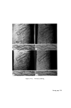
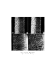
748 JOURNAL OF THE SOCIETY OF COSMETIC CHEMISTS Alternatively, vital dyes may diffuse through the walls of the blood vessels in normal tissue, but fail to colour the tissue in areas of damage where the blood vessels have been destroyed as a result of severe burns (73). Early attempts at using dyes to detect the extent of cutaneous injury due to burns, involved the use of' a modified van Geisen's stain (74) or fluorescein (75). In human studies, vital dyes such as kiton green (76) and disulphine blue (73) injected intravenously, have been found useful in delimiting areas of trauma resulting from severe injury. Tempest (73) considered that the degree of extravasation of the dye was indicative of the severity of. the injury. Although these vital dye methods may be of. value in examining the extent of injury in patients wiith severe burns or crushing, they would appear to have limited application in detecting epidermal damage due to topical applications of. cosmetics, since a subject would be coloured blue or green by this treatment. This is undesirable. Dyes have been employed in animal experiments for detecting the extent of damage due to irritants. Apart from kiton green and disulphine blue as used in clinical studies, pontamine sky blue (77-79), Evan's blue (80) and trypan blue (47, 81) also have been used. In an attempt to quantify the extravasated dye as an index of the tissue damage, Lockett and Jarman (82) compared the intensity of the dye in the skin with a series of comparator cards, Miles and his colleagues (83) measured the diameter of' the zone of increased dye extravasation and expressed this in terms of the 'mean lesion diameter', whereas others made simple visual assessments of the dye intensity in the skin (47, 71, 84). Judah and Willoughby (81), however, believed that none of these methods was accurate enough to quantify an injury and they made extractions of the extravasated dye from tissue samples using standard analytical procedures (78, 80). This is not normally practicable in human studies but when biopsies are taken, these are more profitably examined histologically. The value of' dye extravasation techniques to assess injury which is not evident to the naked eye is questionable since in those studies where dyes have been used, the damage to the skin has been con- siderable and should have been detected unaided (71, 73, 76). Where biopsies can be taken and histological studies conducted, minor degrees of' epidermal damage can be assessed more accurately. Epidermal stripping studies As a means of detecting minor changes in the skin which escape recogni- tion with the naked eye, epidermal stripping may be of value. Anatomical
Purchased for the exclusive use of nofirst nolast (unknown)
From: SCC Media Library & Resource Center (library.scconline.org)