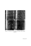
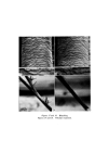
APPRAISAL OF METHODS FOR DETECTING PRIMARY SKIN IRRITANTS 749 studies using this technique date back to the seventeenth century when Marcello Malpighi and others, used warm water to separate the epidermis from underlying tissues in skin samples obtained at autopsy or by biopsy. More recent workers have used ammonia fumes (85) or enzymes (86) to obtain samples of epidermis. However, it is thought that the separation of the epidermis by these methods is likely to result in sufficient damage to obscure the effects due to treatment with an irritant. Another method for separating the epidermis from the dermis relies on the difference in the extensibility of the tissue. By stretching skin samples, the stratum corneum, epidermal cell layer and dermis can be separated (87-89). Although it is unlikely that any useful morphological information could be obtained from tissues prepared in this way, the technique may be suitable for biochemical studies where enzyme assays and analytical studies are contemplated. A technique which has been used for obtaining samples of epidermis from patients, employs blister formation. Blisters induced in the epidermo- dermal region of the skin using chemicals or suction technique, allow an excision of small samples of epidermis to be taken with a minimum of discomfort. 0.25/0 Cantharidin in acetone is capable of raising blisters on the skin of most parts of the body, but on the palms or soles of the feet, 0.55/0 is normally required (90). The extremely toxic nature of this chemical (91, 92) would preclude the use of this technique in routine dermatological studies of cosmetic materials. A method for the induction of blisters using a suction technique was described by Kustala and Mustakallio (93). They used an Angiosterrometer which was applied to the skin for about 3 h, during which time vesiculation and bulla formation occurred in the epidermo-dermal region. Having excised the required sample of epidermis, the remaining tissue of the blister may be pressed back on to its base, re-attachment being complete within 24 h. In this way a minimum of distortion and tissue damage is caused to the excised epidermis and suitable samples are obtained for histological or biochemical studies (94). A drawback with the method con- cerns the inability to induce epidermo-dermal blisters in those skins exhibit- ing pronounced acantholytic changes. In this instance the blisters form in the intra-epidermal region. This observation suggests that the technique is limited in its application to obtaining epidermal samples from skin which is only mildly damaged or is normal. However, the method has been found useful recently in the study of wound healing (95). Adhesive tape has been used for several years to remove the keratinized

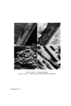
750 JOURNAL OF THE SOCIETY OF COSMETIC CHEMISTS cells from human and animal skins for studying epidermal regeneration (38, 96) and the role of the epidermis in percutaneous absorption (97). Only on a few occasions have workers examined the cells removed for comparative anatomical purposes, or for detecting changes due to the effects of topically applied substances (98). Since Wolf (99, 100) originally described this technique, the method has been modified on several occasions (98, 101, 102). The cells on the surface of the skin are removed using a suitable adhesive tape which is applied using rollers to ensure that complete ad- hesion is achieved. The tape is rapidly removed and with it are the epidermal cells, which can then be stained and examined microscopically. Repeated applications of tape to the skin allow successive layers of cells to be removed down to the Malpighian layer. Although no reports seem to be available to show the value of this technique in examining skins treated topically with irritants, the method is potentially useful for giving information about changes such as hyperkeratosis or parakeratosis, which are frequently present in this type of investigation. Features which make the technique attractive are that it is simple in design and requires little specialized apparatus. The chief disadvantages of the tape stripping technique concern the hairiness of the skin and the irregularity of the layers of the epidermis. With hairy skin, clipping is important and was reported to be preferable to wax depilation or shaving, both of which are liable to result in damage to the superficial layers of the epidermis. The unevenness of the skin, due to the presence of dermatolglyphics and epidermo-dermal ridging, might lead to difficulty in interpretation, since single strippings are likely to contain cells from several layers. This difficulty may be minimized by taking strippings only from small areas of skin. Using a tape stripping technique, it is possible that minor epidermal changes which escape recognition with the naked eye will be detected. It is suitable for use in connection with the patch test, when it is not convenient to take skin biopsies for histological examination. Enzyme studies Histochemical and biochemical studies of enzyme systems have been found useful to detect changes in epidermal metabolism resulting from treatment with hydrocarbons and other agents applied topically. Biochemic- ally, hydroxysteroid dehydrogenase (103), arginase, i.e. L-arginine-amidino- hydrolase (104-107), and protocollagen proline hydroxylase (108), have
Purchased for the exclusive use of nofirst nolast (unknown)
From: SCC Media Library & Resource Center (library.scconline.org)