124 JOURNAL OF THE SOCIETY OF COSMETIC CHEMISTS is altogether inept to speak of "cures." Dandruff can be suppressed but how can a physiologic process be cured (except by death! ). Virtually, nothing is known of the mechanisms, which regulate the rate of epidermal proliferation. Current theories focus on chalones (inhibitors of cell multiplication) and cyclic AMP. Turnover is increased when the concen- tration of the latter is low. No measurements of these regulators have been made on the scalp. We have cursorily looked into the question of whether or not dandruff subjects have increased epidermal turnover in other body areas, the trunk for example. Dandruff subjects give no clinical evidence of increased shedding such as fine scaling, dryness, or roughness. Bearded subjects did not have dan- druff in the beard area. The labeling index was determined on the back and thighs of 4 subjects with severe dandruff. The proportion of DNA synthesiz- ing cells was within the normal range. It is probable that increased des- quamation is limited to the scalp. Finally, it seems unlikely that an animal model of dandruff can be ex- perimentally created. Definitions are all important here. The appearance of dandruff is easily mimicked. Scaling is a nonspecific response to injury. Chem- ical and physical trauma will provoke increased cell turnover and desqua- mation of horny cells in flakes, a dandruff-like condition. Histologically, how- ever, one sees the stigma of tissue injury: inflammatory cells and thickening of the epidermis. In Troller's guinea pig model, the scaling, which followed inoculation of Pityrosporum ovaIe onto skin, anointed with artificial sebum was, in our view, no more than an irritant response to substances liberated from the fatty mixture (8). Rubbing 10 per cent lauric acid in mineral oil daily over the flank of a gninea pig will also induce scaling. Histologic ex- amination immediately shatters the idea that the process is equivalent to dandruff. The telI-tale signs of skin iniury are vividly displayed in the micro- scope. On the other hand, we know of no reason why dandruff should not occur spontaneously in furry animals. Many pet owners think that dogs have dan- druff! IX. Is DANDRUFF A M•LD FORM OF S}mORRm •c DERMATITIS. 9 The statement that dandruff is grade one-half seborrheic dermatitis is trenchant but untrue. These conditions have little in common save scaling. Seborrheic dermatitis is an inflammatory process with a recognizable histolog- ic pattern. As a rule, seborrheic dermatitis will generally show its typical greasy scales on the face and so can easily be recognized. Moreover, unlike dandruff, it is a fluctuating process often being aggravated by emotional stress. There is a widespread, but nonetheless false belief, that dandruff is patchy, involving some portions of the scalp more than others. This idea has resulted

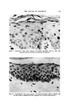
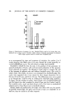
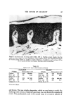
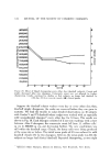
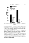
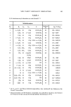
THE NATURE OF DANDRUFF 125 in the practice of dividing the scalp into a number of segments, with each one receiving a separate grade. One scheme calls for separate evaluations of 25 areas, a 20 to 30 min process (4)! Some delineate 9 areas while the "short" method utilizes 4. We compared the corneocyte counts on opposite sides of the scalp in 140 individuals with and without dandruff. The correlation between the two sides was exceptionally high (r--0.843). A phenomenally straight line was pro- duced when the right and left sides were plotted against each other. There was no evidence of asymmetry. Also, by clinical grading, dandruff seems to us to be a uniformly diffused (not patchy) process. Seborrheic dermatitis, on the other hand, presents itself in the form of circumscribed lesions with indistinct borders, just as it does on the skin. It, not dandruff, is patchy. Microorganisms recognize the difference between dandruff and seborrheic dermatitis. While small numbers of S. aureus occassionally can be recovered from dandruff, this potentially virulent organism can be isolated in large num- bers from 20 per cent of the subjects with seborrheic dermatitis (9). S. aureus may comprise as much as one-third of the total flora. Finally, dandruff wanes with age, while seborrheic dermatitis increases. X. HISTOPATHOLOGY No aspect of our study has taken more twists and turns than the views we have held at different times concerning the microscopic changes in dandruff. It will be instructive to review the origins of this confusion. We were satisfied after studying about 50 dandruff specimens that we had a good grasp of the pathologic changes (10). We had examined many normal scalps in the past, and thought we knew the terrain, but our attention then was on the hair follicle, not the epidermis. Naturally, it is quite easy not to see when one is not looking. For a while, we thought that there was in dandruff an increased perivascular infiltrate of mononuclear cells in the upper dermis, a kind of smouldering low grade chronic i•ffiammatory reaction. The idea ap- pealed to us, since at that time, we believed that dandruff was merely mild seborrheic dermatitis. Experts in cutaneous histopathology, who looked at these specimens, agreed with our interpretation, but, of course, they too had no real experience with the anatomy of normal scalps. It is not until one has studied scores of so-called normal scalps that one begins to appreciate the extraordinary individual variations. Many of these show round cells infiltrates to a similar degree. We came to realize that the cuff of adventitial cells, which normally surround small vessels, was greater over the head. The microvascu- lature of the scalp is very richly developed, and mononuclear cells seem to emigrate into the tissues more readily, a sort of slow extravascular circulation. We made the same mistake in studying ache of the face, where, again, nor- mally there are more mononuclear cells patrolling the tissue, creating a spe- cious appearance of low-grade i•ffiammation.
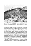
Purchased for the exclusive use of nofirst nolast (unknown)
From: SCC Media Library & Resource Center (library.scconline.org)