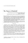
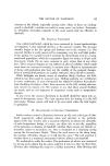
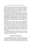
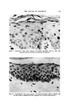
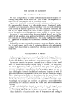
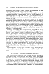
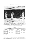
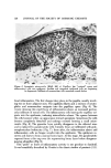
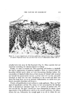
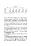
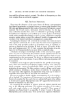
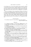
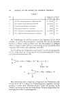
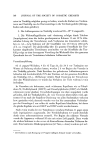
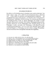
130 JOURNAL OF THE SOCIETY OF COSMETIC CHEMISTS Figure at. Late stage of squame formation (H&E, 265 x). There is a small mound of para- keratotic cells. Underlying epidermis is normal and capillaries are quiescent foinned scales frequently show a solid mass of polys on their surface. The point here is that dandruff scales are heavily impregnated with inflammatory cells and serum and are actually "crusts." It is of more than passing interest that the biochemical changes in dandruff scales have a resemblance to psori- asis, as one might expect, since the dynamics are much the same. Subjects with severe dandruff often coinplain vociferously of scalp itching. Pruritus certainly occurs in nondandruff, also, but is less frequent and less distressing. Itching is, of course, highly variable and perceived differently by individuals. Tentatively, we consider itching to be set off by the squirting capillaries. Inflammatory microfoci in the papillae could readily activate fine sensory nerve fibers. We call particular attention to an artifact, which bewildered us for quite a while. This consisted of small loci of necrotic cells in the upper epidermis. These dead zones stained poorly, were shallow, and had a concave outline. We puzzled over them at length till we perceived that the horny layer over- lying them was ruptured. It finally occurred to us that the horny layer had

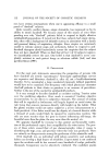
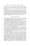
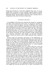
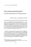
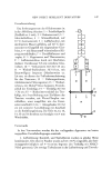
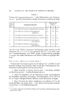
TIlE NATURE OF DANDRUFF 131 Figure 9. A saucer shaved zone of necrosis underlying ruptured horny layer is hallmark of scratch. Fingernail injury has provoked tiny focus of inflammation (H&E, 630 x) actually been torn away by the fingernail (Fig. 9). These necrotic loci are caused by scratching, the natural response to itching! Finally, we nmst re-examine the data regarding cell kinetics in dai•druff. We had no appreciation of squirting capillaries when we scanned slides for thyroidinc labeled nuclei when we were determining labeling indices. Re- examination of dandruff slides showed that clusters of labeled cells occurred just beneath the parakeratotic mounds. Labeled cells were not only unusually abundant in these loci but were distributed in the second and third row above the basal layer, just as in psoriasis. Inflammation, no matter how pro- duced, invariably stimulates cell turnover. Clearly, cell proliferation is not uniform in scalp epidermis. There are "hot" spots of reproductive activity. The values we have presented for the labeling index in dandruff are speciously high, for we counted all labeled cells. We now see that the "hot spots" should have been disregarded to obtain a truer appreciation of the proliferative activity in the normal epidermis. The infiam- nmtory loci are, fortunately, far enough apart so as not to vitiafe the-con-
Purchased for the exclusive use of nofirst nolast (unknown)
From: SCC Media Library & Resource Center (library.scconline.org)