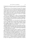
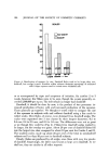
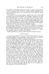
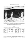
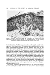
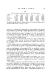
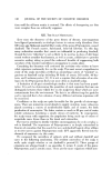
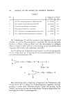
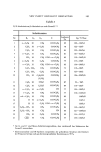
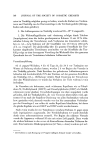
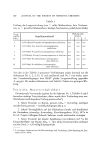
118 JOURNAL OF THE SOCIETY OF COSMETIC CHEMISTS Table I Mitoses Per Thousand Basal Cells Subjects Nondandruff Grades II and III Subjects Dandruff Grades V and VI 14 Mean 14.6 18 Mean 30.6 SD '• ___4.9 SD • ___7.9 :'Standard deviation. Table II Labeling Index (Number of Labeled Cells l•er 100 Basal Cells) S •bjects Nondandruff Grades II and III Subjects Dandruff Grades V and VI l0 Mean 11.0 l0 Mean 17.5 SD -t-2.4 SD ___4.4 ing themselves. Because it is difficult to identify cells in early and late mito- sis, this determination is rather imprecise. Since only a small percentage of basal cells are dividing at a given time, one must scan thousands of cells to keep the experimental error within bounds. Comparisons were made between 14 nondandruff subjects (Grade II and III) and 15 dandruff subjects (14 Grade V and 4 Grade VI). The results are shown in Table I. Even though the values were distributed over a wide range, the mitotic index was about twice as great in the dandruff group (p = 0.01). This result indicates that proliferative activity is increased in dan- druff. This was the first evidence we secured that more cells were being shed from dandruff scalps. It is worth pointing out here that cell turnover on the head region is normally much swifter than on the glabrous skin (6). Another means of estimating cell turnover is to "tag" cells in the reproduc- tive compartment with tritiated thymidine. Those cells which are in the DNA- systhesis phase of the cell cycle will incorporate the radioactive nucleotide into their nuclei. After suitable histologic processing, one can determine the percentage of radio-labeled basal cells (the labeling index). The technique entails a 0.1-ml intradermal injection of 5 to 10 microcuries of tritiated thymi- dine followed by excision biopsy 45 min later. The number of labeled cells per hundred basal cells was determined in 16 subjects without dandruff (Grades II and III) and 19 subjects with Grades V and VI. The results are shown in Table II. Again, the range was great within each group. There can be little doubt, however, that the proportion of DNA-synthesizing cells was about twice as large in dandruff, indicating much faster cell renewal. The rate at which labeled cells move through the epidermis in their journey to the surface (transit time) is another indicator of proliferative activity. To follow cell movement, one delays excising the injected area for a variable number of days. One can then see how far up the tagged cells have migrated

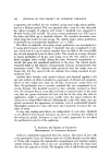
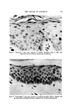
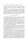
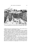
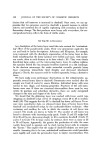
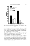
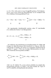
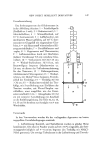
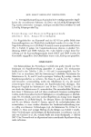
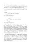
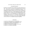
THE NATURE OF DANDRUFF 119 Figure 1. Dandruff, 5 days after injection of tritiated thyroidinc (630 x) cells with labeled nuclei have already reached top of epidermis Figure 2. Nondandruff, 5 days after injection of tritiated thymidine (630 x) labeled cells are fewer than in dandruff and have migrated only to about mid-epidermis
Purchased for the exclusive use of nofirst nolast (unknown)
From: SCC Media Library & Resource Center (library.scconline.org)