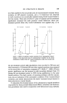
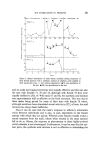
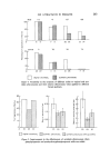
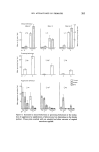
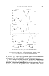
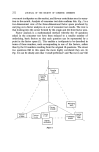
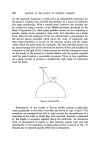
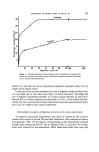
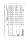
CHEMISTRY OF HUMAN HAIR CUTICLE--Ill 297 but insoluble material remaining after long term trypsin digestion was almost identical in composition to that remaining after 2 h digestion with pronase. (d) Endocuticle subfractions A1 and A2 From our study of the gravimetric course of digestion of whole cuticle with trypsin, two components dissolving at different rates were identified. Since we believe that long term trypsin treatment dissolves the effete cuticle nucleus, it follows that the two new fractions identified are subcomponents of this nucleus. That this is so is in agreement with the morphologic progression of trypsin diges- tion observed with the electron microscope. The component of the cuticle which dissolves in trypsin after 9 h and that which dissolves after treating with trypsin for a further 10 days have been designated endocuticle A1 and A2 respectively (i.e. ENDO A1 and A2). The amino acid analyses of these two subcomponents of the nucleus are shown in columns 5 and 6 of Table I. As would be expected the analyses of A1 and A2 are complementary with respect to the whole ENDO A fraction. In particular A1 contains significantly higher concentrations of aspartic acid, glutamic acid, leucine, tyrosine, phenylalanine and arginine and lower con- centrations of serine, proline, glycine, valine and cystine + cysteic acid than A2. It is also worth pointing out that the analysis for A1 is most extreme from that of whole cuticle. The nuclei of normal viable mammalian cells contain two major types of pro- tein, namely the histone proteins generally characterized by their high basic amino acid content (7), and the non-histone proteins which are relatively rich in acidic amino acids (8). It is difficult to imagine what changes occur to the corresponding proteins from the nuclei of the matrix cells of the hair follicle during the keratin- ization process and certainly their ultimate fate following loss of the nucleic acids of these cells is not known. Perhaps the two fractions A1 and A2 originate from the histone and non-histone proteins of the cuticle cell nucleus modified during the keratinization process. (e) Endocuticle fraction B (ENDO B) From the foregoing it is clear that there is a further fraction of the endocuticle which is not digested by long-term trypsin treatments but which does dissolve in pronase. This component comprises about 15•o by weight of whole cuticle and about 43•o by weight of the endocuticle. The amino acid analysis for this fraction, which we have designated ENDO B, is shown in column 7 of Table I. It is clear from comparison with the ENDO A fraction that ENDO B contains the bulk of the cystine present in the whole endocuticle and that the concentration of aspartic acid is lower.

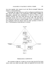
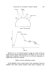
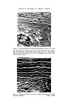
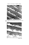
298 JOURNAL OF THE SOCIETY OF COSMETIC CHEMISTS It follows from the foregoing proposals about the origin of ENDO A and its subfractions A1 and A2, that ENDO B probably originates from the non-nuclear cytoplasmic debris of the cuticle cell left over after the formation of the exocuticle, A- and inner-layers. (f) Consideration on the properties and nature of the cuticle cell membrane complex It has been known for some time that single cuticle and cortical cells are apparently liberated into suspension when keratin fibres are treated with solutions of various proteolytic enzymes and it has been generally assumed that this is because the cell membrane complex separating the cells has been degraded (cf. review by Bradbury (9)). From our critical electron microscope examinations of hair sections, treated not only with pronase and trypsin but also with papain/di- thiothreitol (2), we can find no evidence whatsoever for the dissolution of the cuticle cell membrane complex. Even despite extensive enzyme treatments, the full structural integrity and pattern of heavy metal staining of the intercellular membrane glue (5-band) is retained. We therefore believe that the liberation of cuticle cell-like units in the proteolytic enzyme treatment of bulk mammalian keratin fibres is due to digestion along the endocuticle sheet rather than splitting of the cell membrane complex. Further evidence for believing that this is so is that mild mechanical agitation is always necessary to release the cell-like units pre- sumably by rupturing the retaining cell membrane which now only loosely holds the remaining exocuticular segment. A similar situation may also exist in relation to the liberation of cortical cells by proteolytic enzymes with digestion occurring in the intermacrofibrillar matrix, and particularly in that matrix immediately adjacent to the cortical cell membranes, as a necessary prerequisite for subsequent mild mechanical agitation to release cell-like units. The chemical composition of the intercellular membrane cement of the cuticle (5-band) is puzzling. Electron histochemical observations using not only phosphotungstic acid as a stain but also silver/N-acetyl homocysteine thiolactone (10) indicate that this component is rich in free amino groups. If these amino groups were either those of the lysyl groups or the end groups of proteins it is surprising that proteolytic enzymes had no effect on the 5-band. One possibility is that the protein content of the 5-band is small and that the major component is a polysaccharide rich in amino-containing sugar residues. Polysaccharides con- taining vic-diol groupings are certainly present in the cell coat around hair follicle matrix cells and in the intercellular desmosomal plaques between adjacent follicle cells (17). It is not unreasonable to suppose that the polysaccharide coat of the hair follicle cells is retained throughout the keratinization process and, in the case of the cuticle, becomes organized into the layer later identified as the 5-band. Some support for this idea also comes from the fact that we have tentatively identified
Purchased for the exclusive use of nofirst nolast (unknown)
From: SCC Media Library & Resource Center (library.scconline.org)